Copyright
©The Author(s) 2022.
World J Clin Cases. Jul 26, 2022; 10(21): 7474-7482
Published online Jul 26, 2022. doi: 10.12998/wjcc.v10.i21.7474
Published online Jul 26, 2022. doi: 10.12998/wjcc.v10.i21.7474
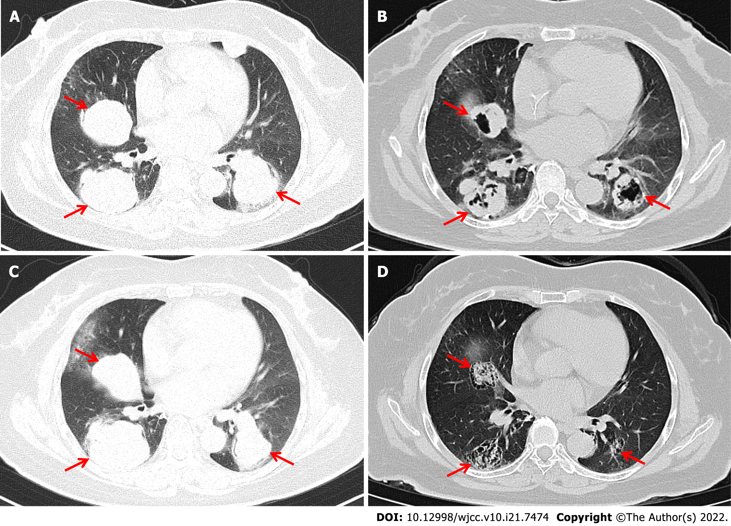
Figure 2 Computed tomography images scans showing changes in lung tumors after combined programmed cell death receptor-1 inhibitor therapy.
Chest computed tomography images showing multiple metastases in bilateral lungs. The three metastases with the largest diameters were selected as target lesions (red arrow). A: The sizes of target lesions before PD-1 inhibitor combination treatment were 47.4 mm, 50.1 mm, and 48.4 mm (6/26/2019); B: The target lesions had regressed in size to 28.9 mm, 37.2 mm, and 32.0 mm after chemotherapy combined with immunotherapy (11/28/2019); C: The target lesions were 57.6 mm, 56.9 mm, and 51.2 mm after 4.4 mo discontinuing treatment (4/16/2020); D: Target lesions had regressed in size to 26.8 mm, 35.8 mm, and 24.3 mm after immunotherapy combined with anti-angiogenesis therapy and massive necrosis was clearly observed (12/23/2020).
- Citation: Zhai CY, Yin LX, Han WD. Programmed cell death-1 inhibitor combination treatment for recurrent proficient mismatch repair/ miscrosatellite-stable type endometrial cancer: A case report. World J Clin Cases 2022; 10(21): 7474-7482
- URL: https://www.wjgnet.com/2307-8960/full/v10/i21/7474.htm
- DOI: https://dx.doi.org/10.12998/wjcc.v10.i21.7474


