Copyright
©The Author(s) 2022.
World J Clin Cases. Jul 26, 2022; 10(21): 7451-7458
Published online Jul 26, 2022. doi: 10.12998/wjcc.v10.i21.7451
Published online Jul 26, 2022. doi: 10.12998/wjcc.v10.i21.7451
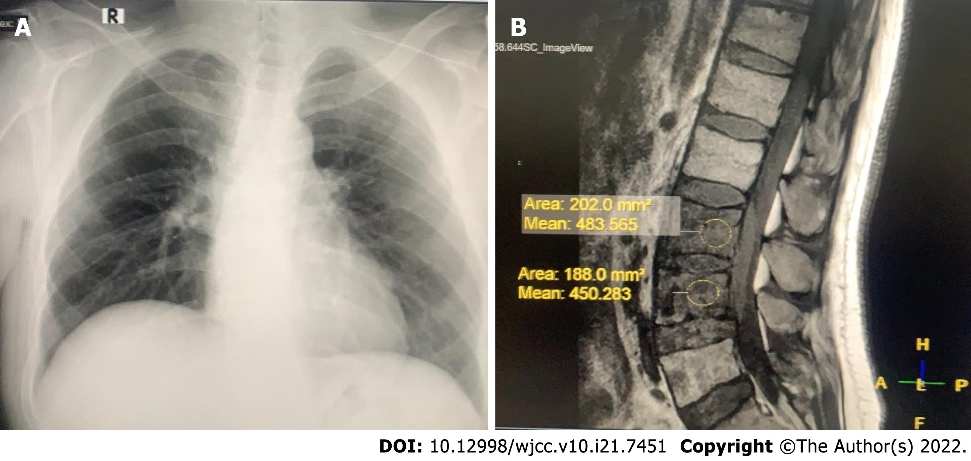
Figure 1 Computed tomography imaging.
A: Chest radiograph of the patient. The lungs were clear, with no masses, granulomas, nodules, consolidation, or collapse visible; B: Magnetic resonance imaging (MRI) of the spine showed a kyphotic thoracic curve, vertebral body destruction at C6, bulging abscess at T9-10, paravertebral abscess formation at L3-4, and abscess extension to the anterior spinal canal.
- Citation: Novita BD, Muliono AC, Wijaya S, Theodora I, Tjahjono Y, Supit VD, Willianto VM. Managing spondylitis tuberculosis in a patient with underlying diabetes and hypothyroidism: A case report. World J Clin Cases 2022; 10(21): 7451-7458
- URL: https://www.wjgnet.com/2307-8960/full/v10/i21/7451.htm
- DOI: https://dx.doi.org/10.12998/wjcc.v10.i21.7451


