Copyright
©The Author(s) 2022.
World J Clin Cases. Jul 26, 2022; 10(21): 7445-7450
Published online Jul 26, 2022. doi: 10.12998/wjcc.v10.i21.7445
Published online Jul 26, 2022. doi: 10.12998/wjcc.v10.i21.7445
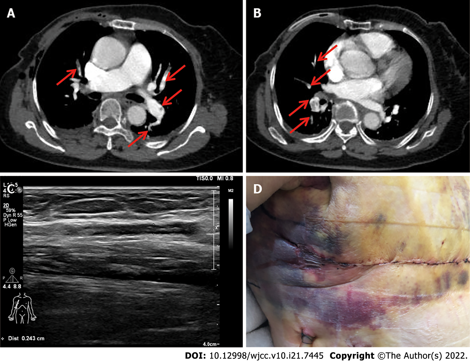
Figure 2 Computed tomography pulmonary angiograms, vascular Doppler ultrasound, and surgical wound.
A: Computed tomography angiography demonstrated massive thrombosis in the segmental arteries of the right upper lobe, segmental arteries of the left upper lobe, and segmental arteries of the left lower lobe (red arrow heads); B: CT angiography revealed filling defects in the segmental arteries of the middle lobe and segmental arteries of the right lower lobe (red arrow heads); C: Doppler ultrasound revealed a venous thrombosis in the right vena basilica; D: Subcutaneous ecchymosis caused by anticoagulant therapy.
- Citation: Duan Y, Wang GL, Guo X, Yang LL, Tian FG. Acute pulmonary embolism originating from upper limb venous thrombosis following breast cancer surgery: Two case reports. World J Clin Cases 2022; 10(21): 7445-7450
- URL: https://www.wjgnet.com/2307-8960/full/v10/i21/7445.htm
- DOI: https://dx.doi.org/10.12998/wjcc.v10.i21.7445


