Copyright
©The Author(s) 2022.
World J Clin Cases. Jul 26, 2022; 10(21): 7445-7450
Published online Jul 26, 2022. doi: 10.12998/wjcc.v10.i21.7445
Published online Jul 26, 2022. doi: 10.12998/wjcc.v10.i21.7445
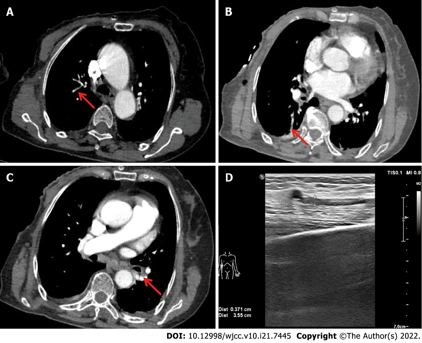
Figure 1 Computed tomography pulmonary angiograms and vascular Doppler ultrasound.
A-C: Computed tomography pulmonary angiograms showing extensive bilateral filling defects in the segmental arteries of the right upper lobe (A), segmental arteries of the right lower lobe (B), and segmental arteries of the left lower lobe (C); D: Doppler ultrasound revealed a deep venous thrombosis in the brachial vein of the right upper limb.
- Citation: Duan Y, Wang GL, Guo X, Yang LL, Tian FG. Acute pulmonary embolism originating from upper limb venous thrombosis following breast cancer surgery: Two case reports. World J Clin Cases 2022; 10(21): 7445-7450
- URL: https://www.wjgnet.com/2307-8960/full/v10/i21/7445.htm
- DOI: https://dx.doi.org/10.12998/wjcc.v10.i21.7445


