Copyright
©The Author(s) 2022.
World J Clin Cases. Jul 26, 2022; 10(21): 7438-7444
Published online Jul 26, 2022. doi: 10.12998/wjcc.v10.i21.7438
Published online Jul 26, 2022. doi: 10.12998/wjcc.v10.i21.7438
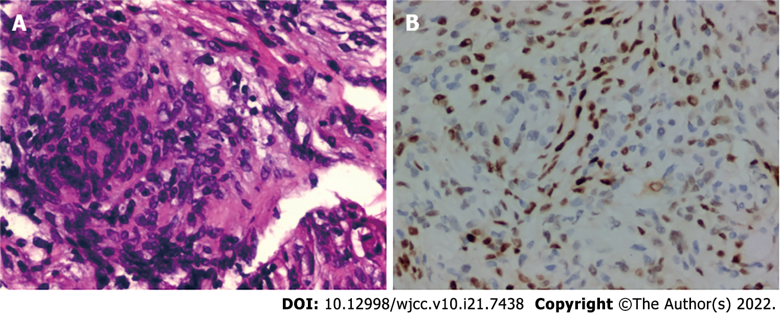
Figure 3 Postoperative histopathology.
A: Hematoxylin and eosin staining; original magnification, × 200: Microscopically, the tissue demonstrates spindle-shaped tumor cells and characteristic diffused arrangement in sheets with multiple gravel structures; B: Estimated proliferation index of 5% stained by Ki67 (Immunohistochemical staining, original magnification, × 200). Considering the H&E results, findings were consistent with meningioma.
- Citation: Wang A, Zhang X, Sun KK, Li C, Song ZM, Sun T, Wang F. Deep Sylvian fissure meningiomas: A case report. World J Clin Cases 2022; 10(21): 7438-7444
- URL: https://www.wjgnet.com/2307-8960/full/v10/i21/7438.htm
- DOI: https://dx.doi.org/10.12998/wjcc.v10.i21.7438


