Copyright
©The Author(s) 2022.
World J Clin Cases. Jul 26, 2022; 10(21): 7415-7421
Published online Jul 26, 2022. doi: 10.12998/wjcc.v10.i21.7415
Published online Jul 26, 2022. doi: 10.12998/wjcc.v10.i21.7415
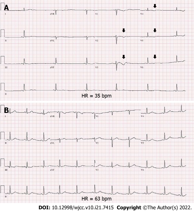
Figure 2 Electrocardiogram of the patient.
A: Initial electrocardiogram (ECG) reveals marked sinus bradycardia (heart rate of 35 bpm) with flattening and inversion of T-waves (black arrows), mainly in the precordial leads (V1–V5); B: Follow-up ECG showing normalized heart rate and T-wave configuration after steroid discontinuation. ECG: Electrocardiogram; MRI: Magnetic resonance imaging; FLAIR: Fluid-attenuated inversion recovery image; mPD: Methylprednisolone; bpm: Beats per minute.
- Citation: Sohn SY, Kim SY, Joo IS. Corticosteroid-induced bradycardia in multiple sclerosis and maturity-onset diabetes of the young due to hepatocyte nuclear factor 4-alpha mutation: A case report. World J Clin Cases 2022; 10(21): 7415-7421
- URL: https://www.wjgnet.com/2307-8960/full/v10/i21/7415.htm
- DOI: https://dx.doi.org/10.12998/wjcc.v10.i21.7415


