Copyright
©The Author(s) 2022.
World J Clin Cases. Jul 26, 2022; 10(21): 7397-7408
Published online Jul 26, 2022. doi: 10.12998/wjcc.v10.i21.7397
Published online Jul 26, 2022. doi: 10.12998/wjcc.v10.i21.7397
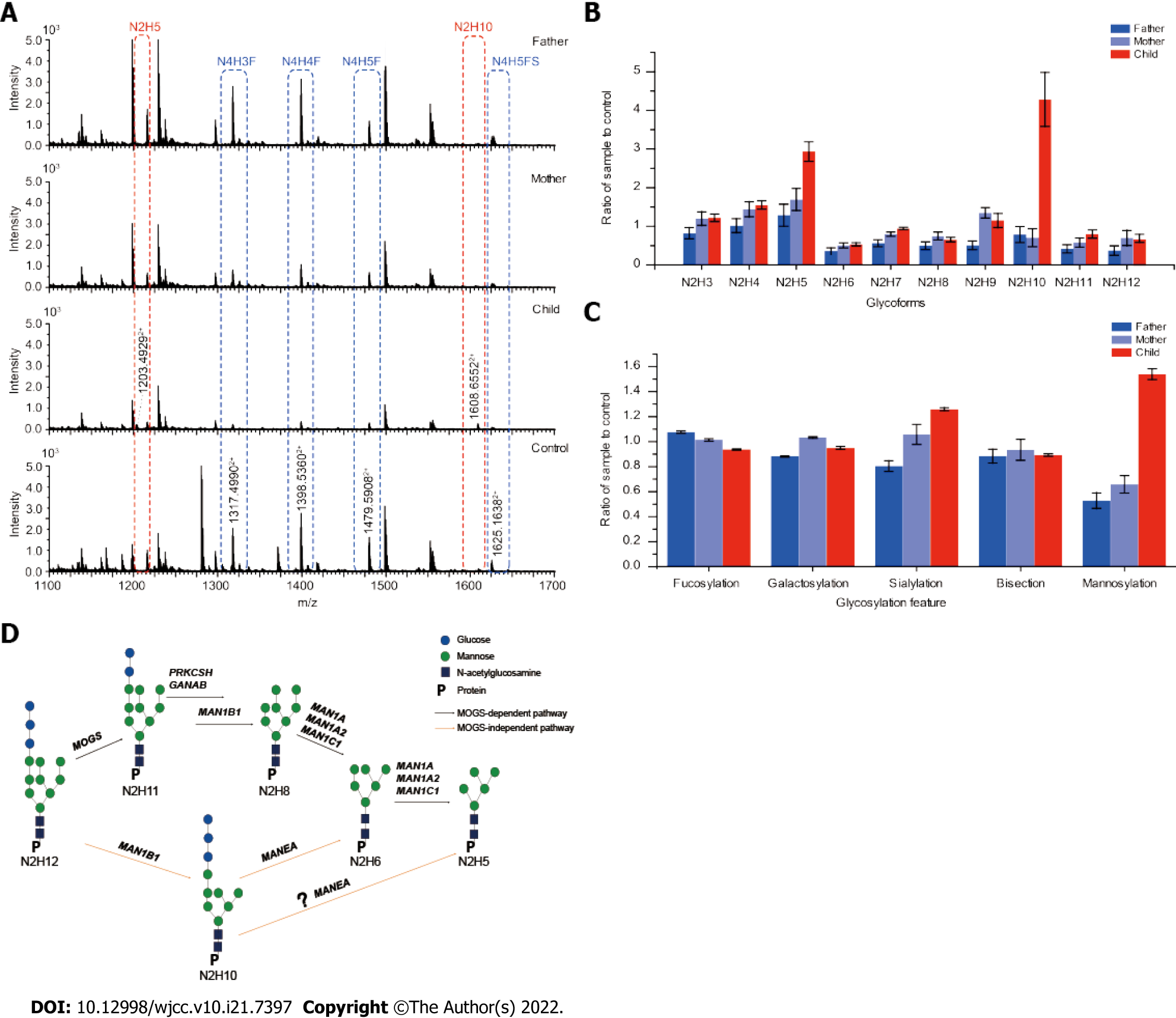
Figure 2 Abnormal glycoforms detected on immunoglobulin G1 in the serum of the younger sister and proposed compensatory pathway in mannosyl-oligosaccharide glucosidase-congenital disorder of glycosylation.
A: Representative mass spectrums generated from the younger sister, parents, and pooled healthy controls by nano-electrospray ionization quadrupole time-of-flight mass spectrometry; B: N2 serials of oligosaccharides on immunoglobulin G1 (IgG1) normalized by the control. Abnormal enrichment of N2H10 and N2H5 were detected; C: Fractions of IgG1 with fucosylation, galactosylation, sialylation, mannosylation, and bisection showing significantly high levels mannosylation and sialylation in the patient sample; D: Proposed mannosyl-oligosaccharide glucosidase-independent pathway leading to increased levels of N2H10 and N2H5 (M5) in vivo. N: N-acetylglucosamine and/or N-acetylgalactosamine; H, hexaose (galactose, glucose, or mannose); GANAB: α-Subunit of glucosidase II; PRKCSH: β-Subunit of glucosidase II; MAN1B1: Endoplasmic reticulum mannosyl-oligosaccharide α-1,2-mannosidase; MAN1A, MAN1A2, and MAN1C1: Mannosyl-oligosaccharide α-1,2-mannosidase; MANEA: Endo-alpha-1,2-mannosidase.
- Citation: Abuduxikuer K, Wang L, Zou L, Cao CY, Yu L, Guo HM, Liang XM, Wang JS, Chen L. Updated clinical and glycomic features of mannosyl-oligosaccharide glucosidase deficiency: Two case reports. World J Clin Cases 2022; 10(21): 7397-7408
- URL: https://www.wjgnet.com/2307-8960/full/v10/i21/7397.htm
- DOI: https://dx.doi.org/10.12998/wjcc.v10.i21.7397


