Copyright
©The Author(s) 2022.
World J Clin Cases. Jul 16, 2022; 10(20): 7178-7183
Published online Jul 16, 2022. doi: 10.12998/wjcc.v10.i20.7178
Published online Jul 16, 2022. doi: 10.12998/wjcc.v10.i20.7178
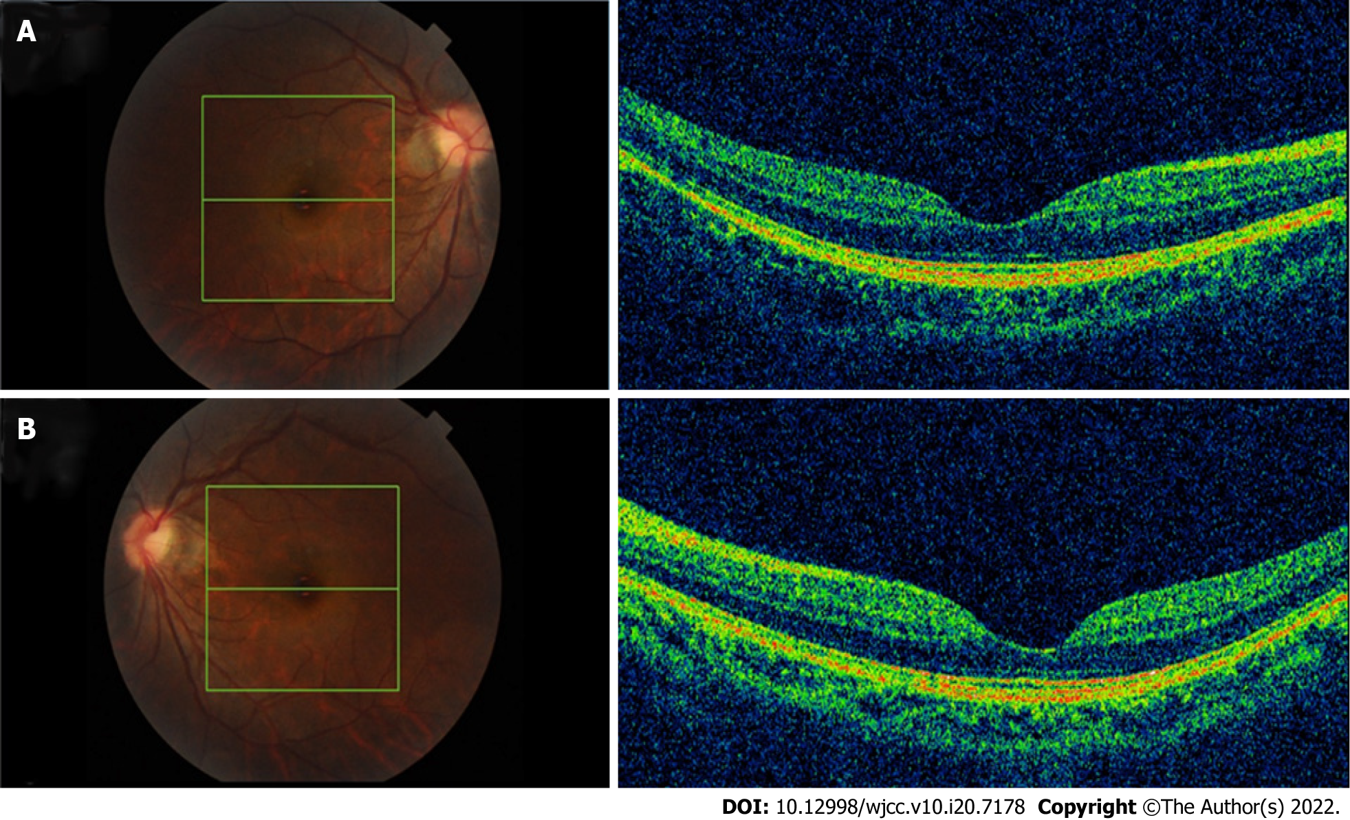
Figure 2 Optical coherence tomography examination of both eyes before phakic intraocular lens surgery revealed a normal macula, without posterior vitreous detachment, posterior staphyloma, or foveoschisis.
A: Right eye; B: Left eye.
- Citation: Li XJ, Duan JL, Ma JX, Shang QL. Macular hole following phakic intraocular lens implantation: A case report. World J Clin Cases 2022; 10(20): 7178-7183
- URL: https://www.wjgnet.com/2307-8960/full/v10/i20/7178.htm
- DOI: https://dx.doi.org/10.12998/wjcc.v10.i20.7178


