Copyright
©The Author(s) 2022.
World J Clin Cases. Jul 16, 2022; 10(20): 7153-7162
Published online Jul 16, 2022. doi: 10.12998/wjcc.v10.i20.7153
Published online Jul 16, 2022. doi: 10.12998/wjcc.v10.i20.7153
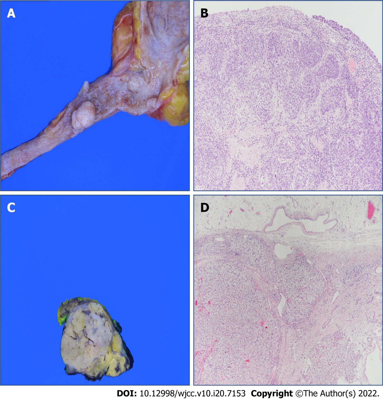
Figure 5 Pathologic findings.
A: The specimen shows multiple protruding nodular masses in left proximal ureter, measuring 1.7 cm × 1.0 cm in the largest one; B: High-grade urothelial carcinoma in the left proximal ureter, shows invasion to the muscular layer. [hematoxylin-eosin staining (H&E), 100 ×]; C: The cut surface reveals a 3.8 cm × 3.2 cm yellowish, well-demarcated solid tumor in right kidney; D: Renal cell carcinoma of clear cell type with Fuhrman nuclear Grade 3 in right kidney, shows invasion to perinephric tissue (H&E, 40 ×).
- Citation: Yun JK, Kim SH, Kim WB, Kim HK, Lee SW. Simultaneous robot-assisted approach in a super-elderly patient with urothelial carcinoma and synchronous contralateral renal cell carcinoma: A case report. World J Clin Cases 2022; 10(20): 7153-7162
- URL: https://www.wjgnet.com/2307-8960/full/v10/i20/7153.htm
- DOI: https://dx.doi.org/10.12998/wjcc.v10.i20.7153


