Copyright
©The Author(s) 2022.
World J Clin Cases. Jul 16, 2022; 10(20): 7153-7162
Published online Jul 16, 2022. doi: 10.12998/wjcc.v10.i20.7153
Published online Jul 16, 2022. doi: 10.12998/wjcc.v10.i20.7153
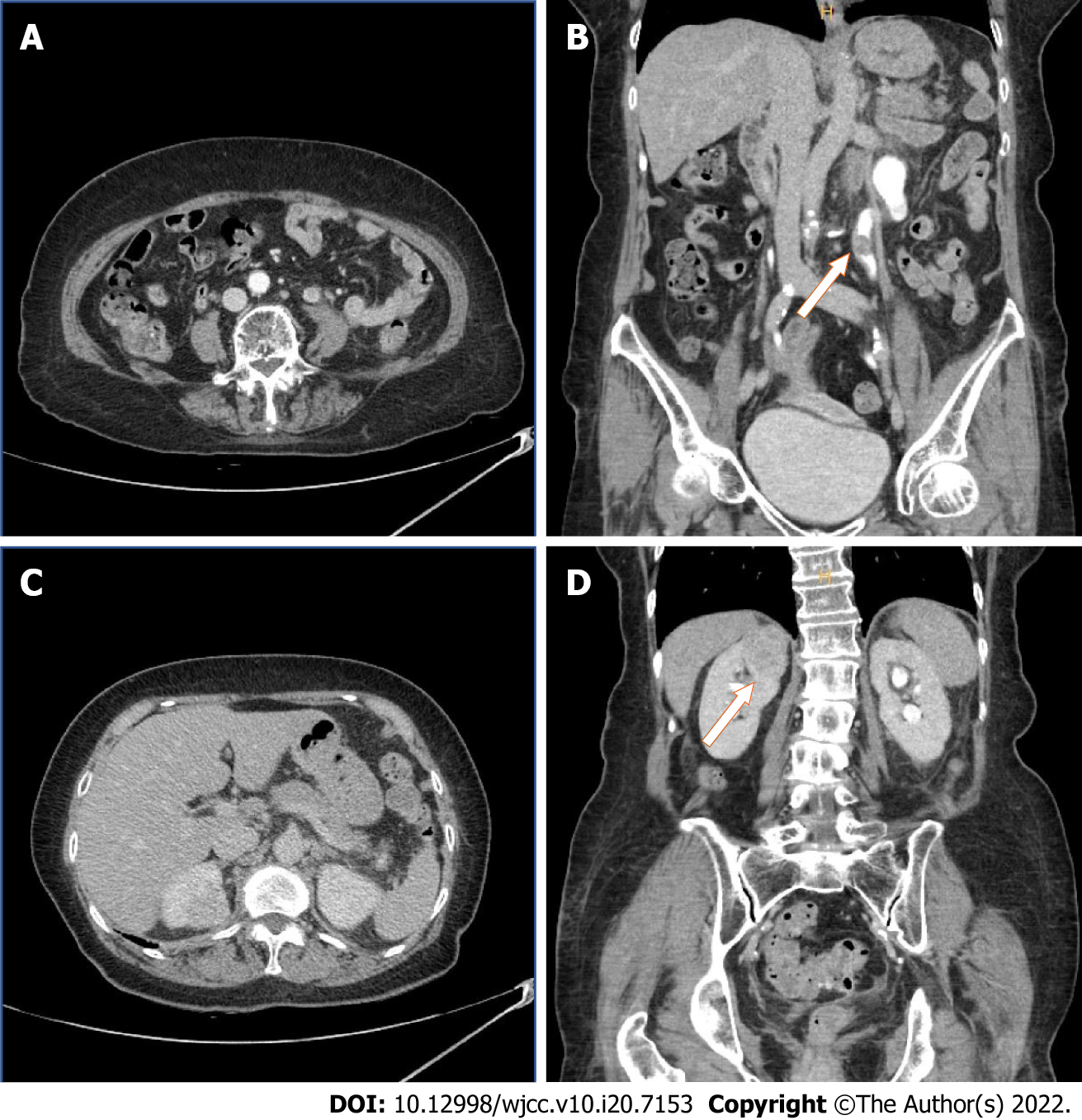
Figure 2 After admission, computed tomography urography was performed to obtain exact image of tumor distribution.
A 1.6 cm-sized mass was observed in the left proximal ureter’s distal portion with suspicious periureteral soft tissue invasion. No regional lymph node enlargement was founded. A: Axial; B: Coronal. 3.1 cm-sized lobulation, relatively homogeneous enhancing mass was observed. Intratumoral cyst with focal oval low density area was founded. Most likely renal cell carcinoma.
- Citation: Yun JK, Kim SH, Kim WB, Kim HK, Lee SW. Simultaneous robot-assisted approach in a super-elderly patient with urothelial carcinoma and synchronous contralateral renal cell carcinoma: A case report. World J Clin Cases 2022; 10(20): 7153-7162
- URL: https://www.wjgnet.com/2307-8960/full/v10/i20/7153.htm
- DOI: https://dx.doi.org/10.12998/wjcc.v10.i20.7153


