Copyright
©The Author(s) 2022.
World J Clin Cases. Jul 16, 2022; 10(20): 7153-7162
Published online Jul 16, 2022. doi: 10.12998/wjcc.v10.i20.7153
Published online Jul 16, 2022. doi: 10.12998/wjcc.v10.i20.7153
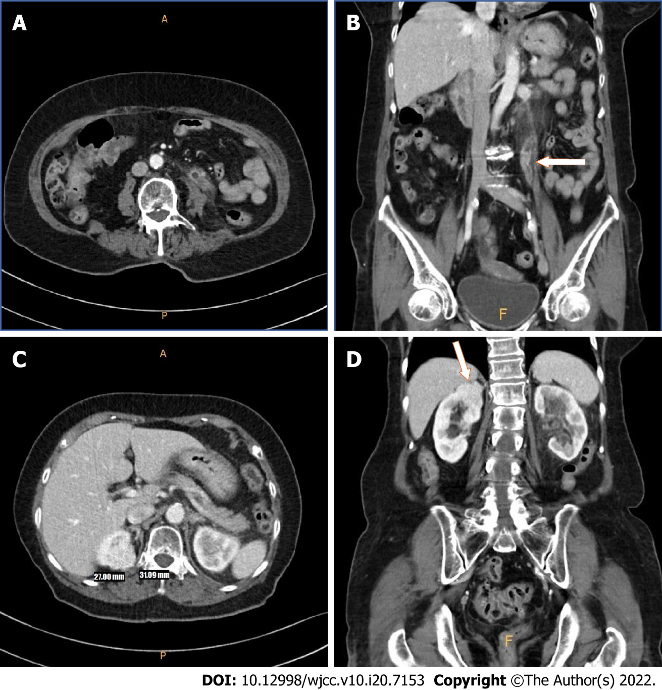
Figure 1 Computed tomography.
Pre-operative computed tomography showed proximal segmental enhancing mass with eccentric left ureteral wall thickness. suggestive of urothelial tumor. A: Axial; B: Coronal. In the upper pole of the right kidney, a 3 cm-sized enhancing mass was identified. Suspected of renal cell carcinoma. No direct invasion was identified; C: Axial; D: Coronal.
- Citation: Yun JK, Kim SH, Kim WB, Kim HK, Lee SW. Simultaneous robot-assisted approach in a super-elderly patient with urothelial carcinoma and synchronous contralateral renal cell carcinoma: A case report. World J Clin Cases 2022; 10(20): 7153-7162
- URL: https://www.wjgnet.com/2307-8960/full/v10/i20/7153.htm
- DOI: https://dx.doi.org/10.12998/wjcc.v10.i20.7153


