Copyright
©The Author(s) 2022.
World J Clin Cases. Jul 16, 2022; 10(20): 7124-7129
Published online Jul 16, 2022. doi: 10.12998/wjcc.v10.i20.7124
Published online Jul 16, 2022. doi: 10.12998/wjcc.v10.i20.7124
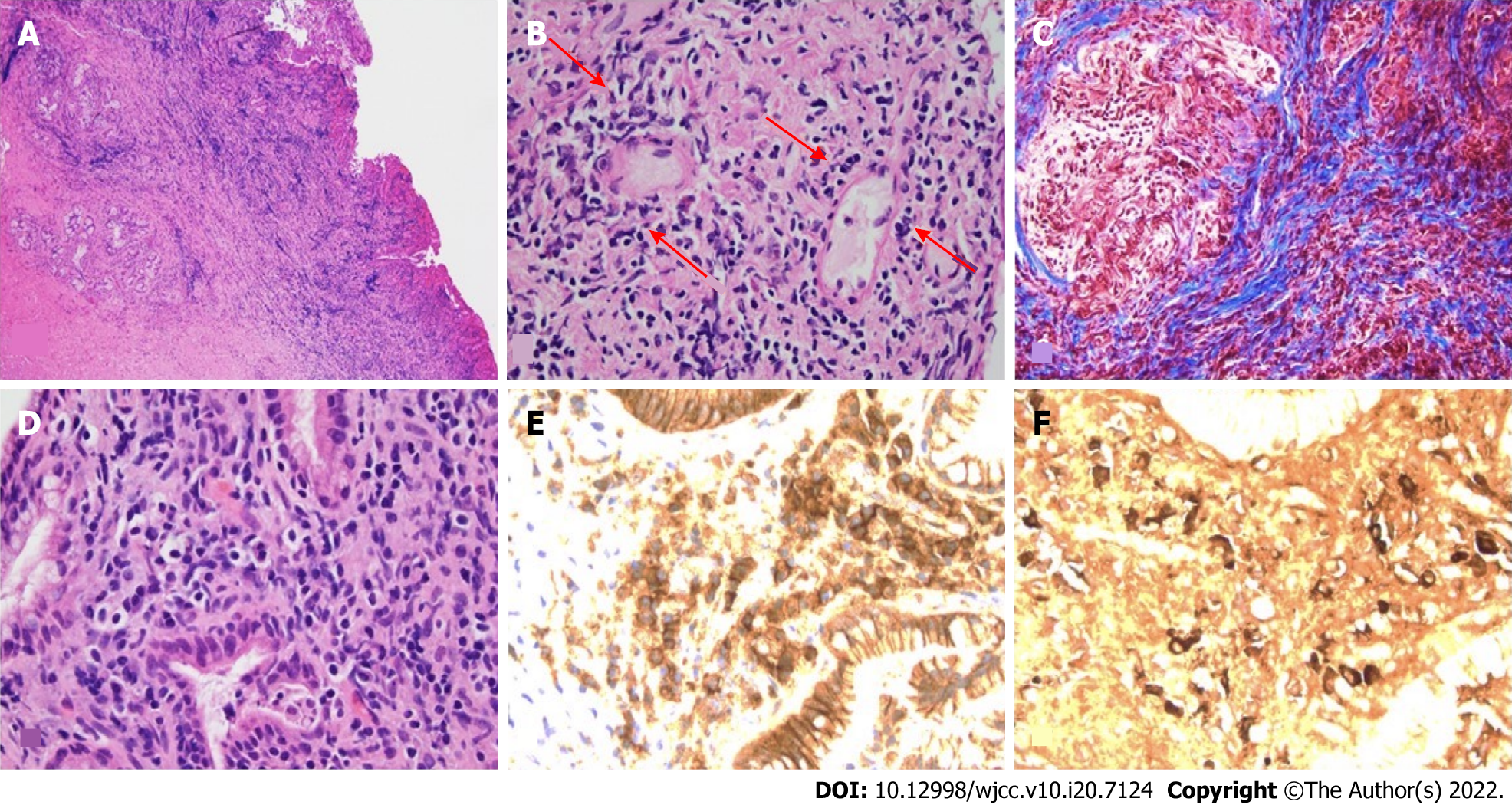
Figure 2 Hematoxylin and eosin stained.
A: Low power hematoxylin and eosin (H&E) showing marked chronic mixed cell infiltrates and fibrosis within epithelial and subepithelial region; B: Medium power H&E microscopy showing venules flanked by lymphocytes consistent with lymphocytic phlebitis (arrows); C: Medium power trichrome microscopy showing Prussian blue trichome staining of fibrosis with storiform pattern; D: High power H&E microscopy showing plasma call predominant inflammatory cell infiltrates amidst biliary glands; E: High power view of same area in D, showing numerous plasma cells highlighted with dark brown signal by CD138 immunohistochemistry antibody, a plasma cell biomarker; F: High power view of same area in D, showing numerous immunoglobulin G4 (IgG4) positive plasma cells highlighted with dark brown signals by IgG4 immunohistochemistry antibody.
- Citation: Agrawal R, Guzman G, Karimi S, Giulianotti PC, Lora AJM, Jain S, Khan M, Boulay BR, Chen Y. Immunoglobulin G4 associated autoimmune cholangitis and pancreatitis following the administration of nivolumab: A case report. World J Clin Cases 2022; 10(20): 7124-7129
- URL: https://www.wjgnet.com/2307-8960/full/v10/i20/7124.htm
- DOI: https://dx.doi.org/10.12998/wjcc.v10.i20.7124


