Copyright
©The Author(s) 2022.
World J Clin Cases. Jul 16, 2022; 10(20): 7082-7089
Published online Jul 16, 2022. doi: 10.12998/wjcc.v10.i20.7082
Published online Jul 16, 2022. doi: 10.12998/wjcc.v10.i20.7082
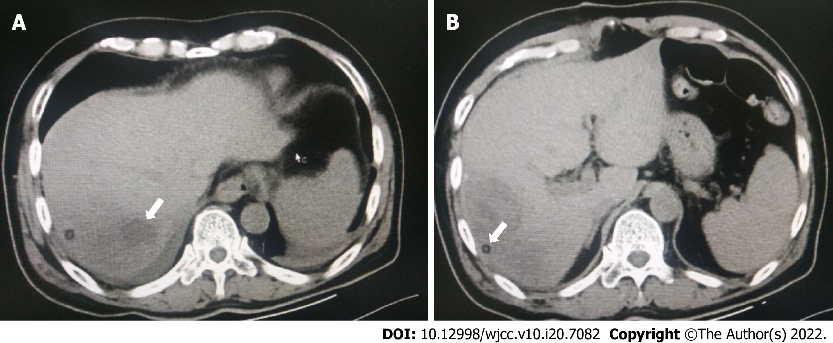
Figure 4 Radiographs taken 3 d after the procedure.
A: The intrahepatic low-attenuation lesion was filled with platelet-rich plasma (white arrow); B: The drainage tube (white arrow) was visible in the abscess cavity.
- Citation: Wang JH, Gao ZH, Qian HL, Li JS, Ji HM, Da MX. Treatment of pyogenic liver abscess by surgical incision and drainage combined with platelet-rich plasma: A case report. World J Clin Cases 2022; 10(20): 7082-7089
- URL: https://www.wjgnet.com/2307-8960/full/v10/i20/7082.htm
- DOI: https://dx.doi.org/10.12998/wjcc.v10.i20.7082


