Copyright
©The Author(s) 2022.
World J Clin Cases. Jul 16, 2022; 10(20): 7045-7053
Published online Jul 16, 2022. doi: 10.12998/wjcc.v10.i20.7045
Published online Jul 16, 2022. doi: 10.12998/wjcc.v10.i20.7045
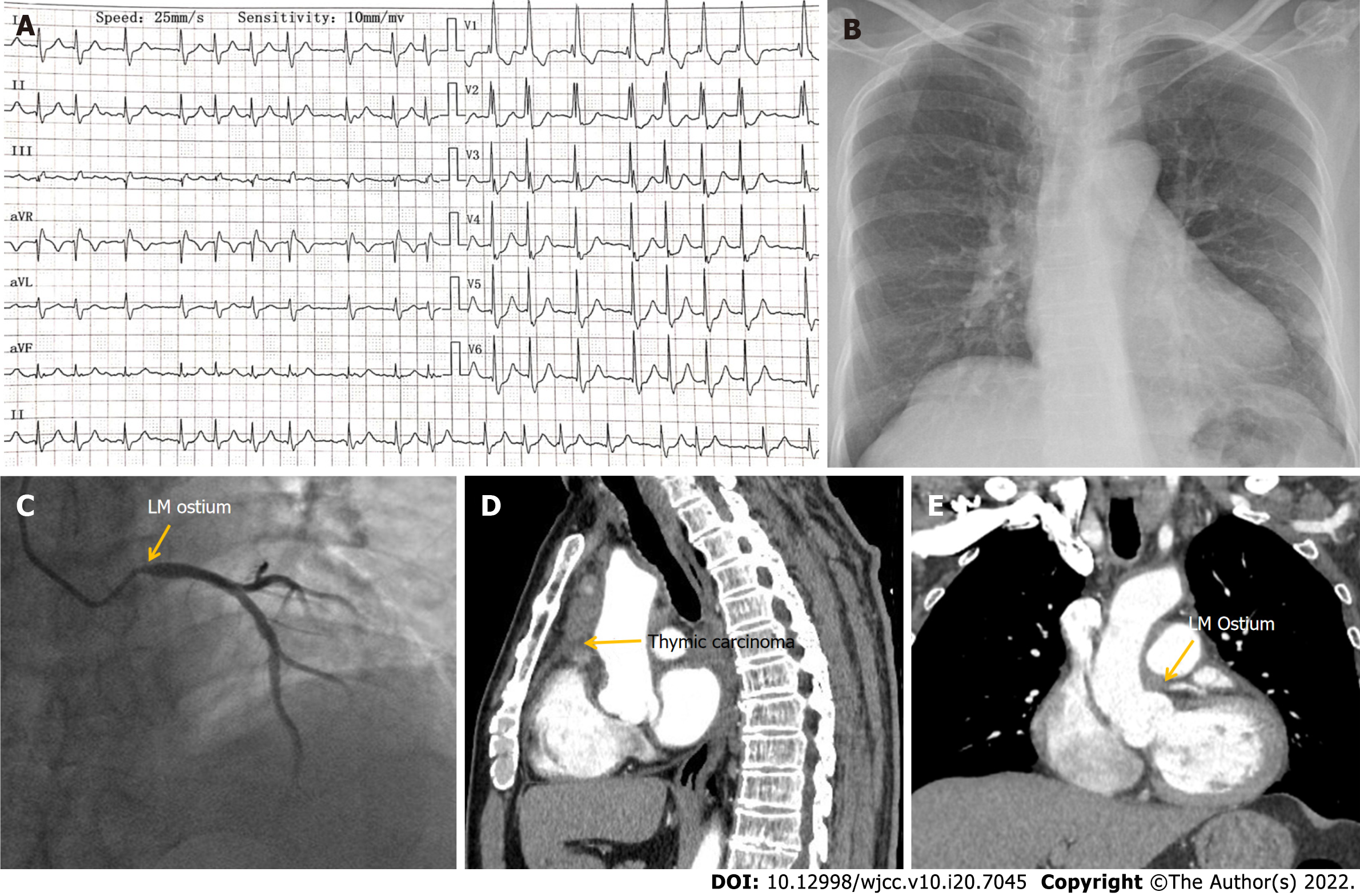
Figure 1 Electrocardiography and imaging manifestations of Case 1.
A: Electrocardiography showed atrial fibrillation, complete right bundle branch block and depressed ST segment of 0.1–0.3 mV in leads V1-V6; B: Chest X-ray was normal; C: Coronary angiography showed severe stenosis in the left main coronary ostium; D: Chest computed tomography (CT) showed thymic carcinoma involving the middle mediastinum; E: Chest CT showed thymic carcinoma involving the middle mediastinum, which also invaded the left main coronary ostium. LM: Left main.
- Citation: Liu XP, Wang HJ, Gao JL, Ma GL, Xu XY, Ji LN, He RX, Qi BYE, Wang LC, Li CQ, Zhang YJ, Feng YB. Secondary coronary artery ostial lesions: Three case reports. World J Clin Cases 2022; 10(20): 7045-7053
- URL: https://www.wjgnet.com/2307-8960/full/v10/i20/7045.htm
- DOI: https://dx.doi.org/10.12998/wjcc.v10.i20.7045


