Copyright
©The Author(s) 2022.
World J Clin Cases. Jul 16, 2022; 10(20): 7013-7019
Published online Jul 16, 2022. doi: 10.12998/wjcc.v10.i20.7013
Published online Jul 16, 2022. doi: 10.12998/wjcc.v10.i20.7013
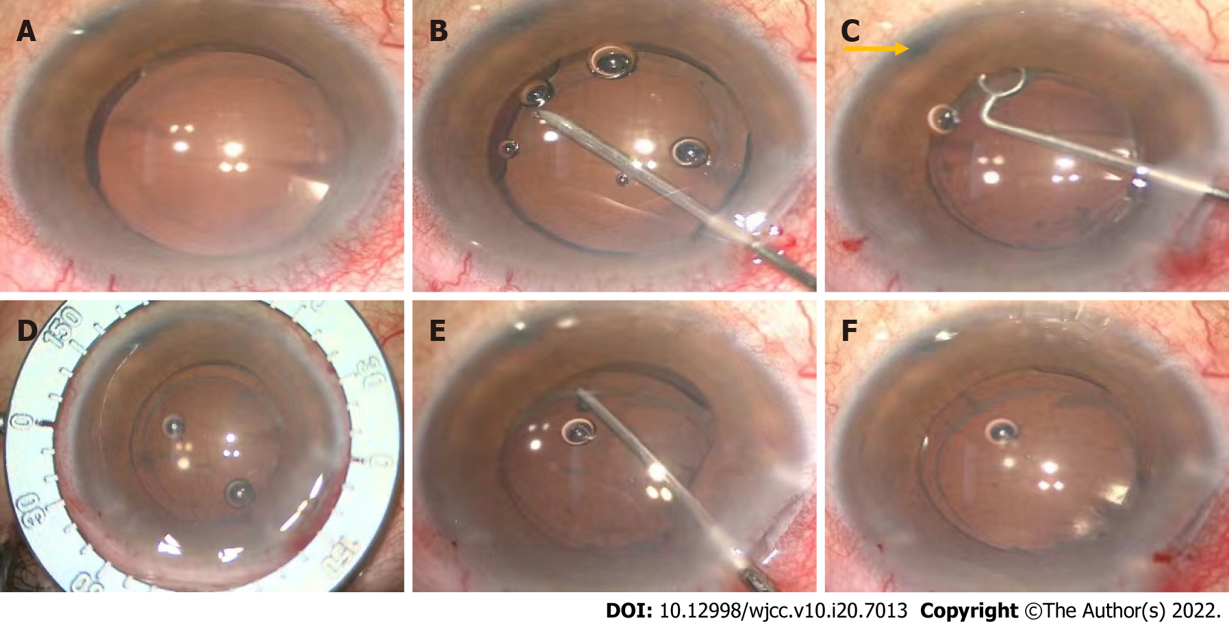
Figure 2 Intraoperative images of the rotating lens.
A: Preoperatively, the light projection of the microscope coincides with the center of the intraocular lens (IOL); B: Separating capsulorhexis opening with a needle; C: Polishing anterior capsule. The laser hole (yellow arrow) after peripheral iridectomy is clearly visible; D: Locating the position of the IOL after rotation with a ring manually; E: Adjusting the position of the IOL with an IOL hook; F: The light projection of the microscope is on the central point of the IOL after rotation.
- Citation: Fan C, Zhou Y, Jiang J. Secondary positioning of rotationally asymmetric refractive multifocal intraocular lens in a patient with glaucoma: A case report. World J Clin Cases 2022; 10(20): 7013-7019
- URL: https://www.wjgnet.com/2307-8960/full/v10/i20/7013.htm
- DOI: https://dx.doi.org/10.12998/wjcc.v10.i20.7013


