Copyright
©The Author(s) 2022.
World J Clin Cases. Jul 16, 2022; 10(20): 6991-6998
Published online Jul 16, 2022. doi: 10.12998/wjcc.v10.i20.6991
Published online Jul 16, 2022. doi: 10.12998/wjcc.v10.i20.6991
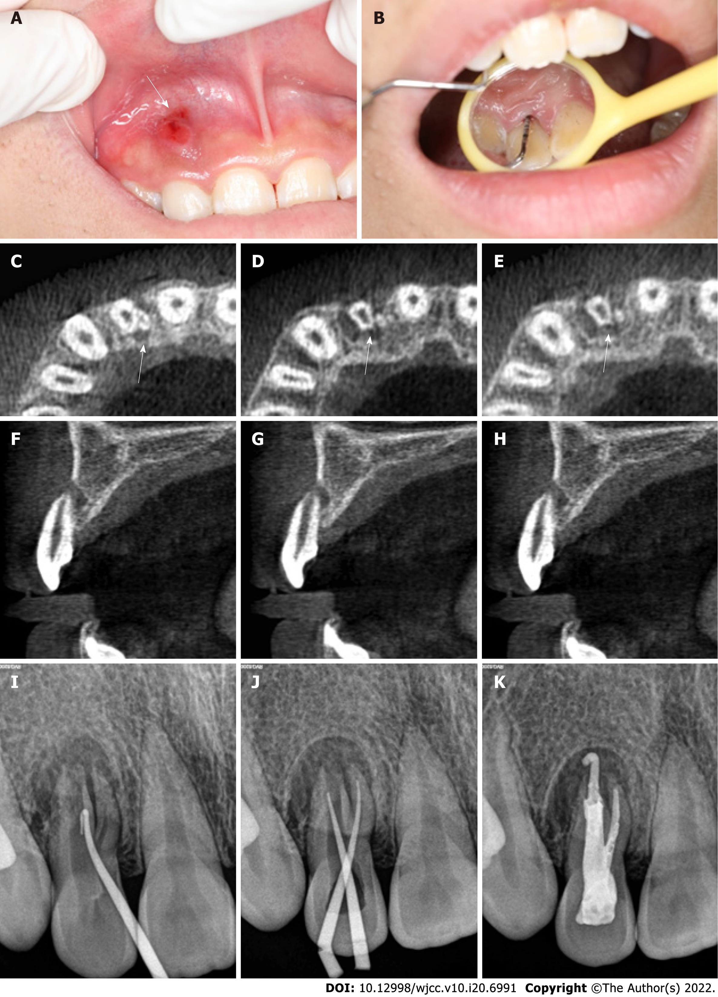
Figure 1 Oral examination and imaging examination before surgery.
A: Visual examination revealed a draining sinus tract on the labial gingival surface associated with the maxillary right lateral incisor; B: Periodontal probing depth of the involved tooth was 13 mm; C-H: Cone-beam computed tomography showed that the maxillary right lateral incisor had two roots with a large area of periradicular radiolucency; I: Preoperative radiography showing that gutta percha was used to trace the sinus tract; J: Intraoral radiography immediately after root canal preparation; K: Intraoral radiography immediately after root canal therapy.
- Citation: Tan D, Li ST, Feng H, Wang ZC, Wen C, Nie MH. Intentional replantation combined root resection therapy for the treatment of type III radicular groove with two roots: A case report. World J Clin Cases 2022; 10(20): 6991-6998
- URL: https://www.wjgnet.com/2307-8960/full/v10/i20/6991.htm
- DOI: https://dx.doi.org/10.12998/wjcc.v10.i20.6991


