Copyright
©The Author(s) 2022.
World J Clin Cases. Jul 16, 2022; 10(20): 6981-6990
Published online Jul 16, 2022. doi: 10.12998/wjcc.v10.i20.6981
Published online Jul 16, 2022. doi: 10.12998/wjcc.v10.i20.6981
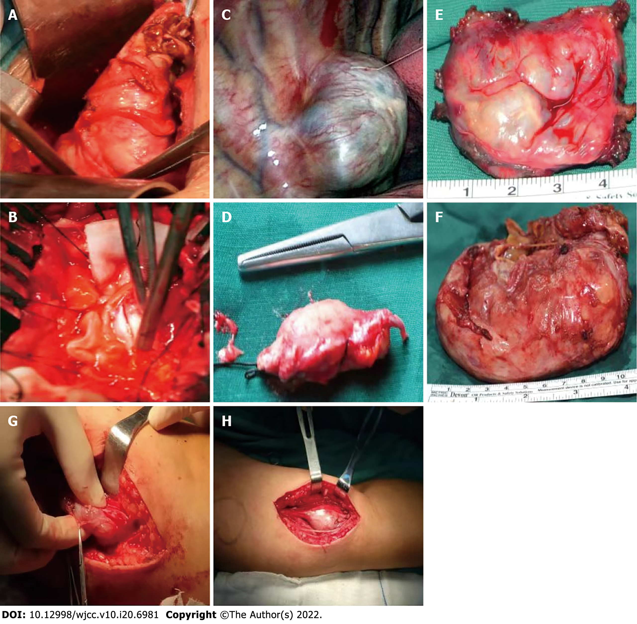
Figure 5 Intraoperative images.
A-F: Intraoperative images from previous surgeries. A-C: Schwannoma exposed during the operation; D: Schwannoma tissue removed during lumbar spinal surgery; E: Schwannoma tissue removed during thoracic spinal surgery; F: Large schwannoma tissue specimen removed during pelvic surgery. G-H: Intraoperative images from recent surgeries, schwannoma exposed in the left forearm of this patient.
- Citation: Li K, Liu SJ, Wang HB, Yin CY, Huang YS, Guo WT. Schwannomatosis patient who was followed up for fifteen years: A case report. World J Clin Cases 2022; 10(20): 6981-6990
- URL: https://www.wjgnet.com/2307-8960/full/v10/i20/6981.htm
- DOI: https://dx.doi.org/10.12998/wjcc.v10.i20.6981


