Copyright
©The Author(s) 2022.
World J Clin Cases. Jan 14, 2022; 10(2): 725-732
Published online Jan 14, 2022. doi: 10.12998/wjcc.v10.i2.725
Published online Jan 14, 2022. doi: 10.12998/wjcc.v10.i2.725
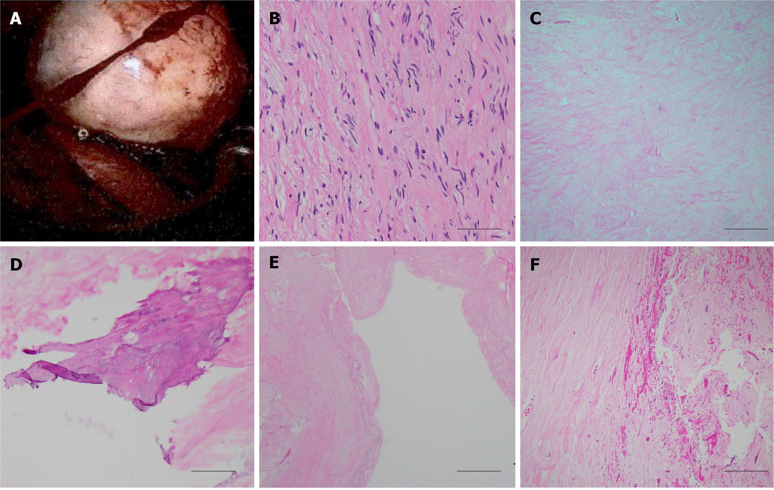
Figure 3 Histological examination of the spinal tumor.
A: Intraoperative image of the posterior mediastinal tumor demonstrated well-defined border, which was pathologically proved to be neurogenic tumor; B-F: A histological image of the neurofibroma showing bland spindle cells with wavy nuclei and pale eosinophilic cytoplasm (scale bar 50 μm) (B), secondary degeneration of hyalinization (scale bar 200 μm) (C), calcification (scale bar 100 μm) (D), cyst (scale bar 500 μm) (E), and hemorrhage (scale bar 200 μm) (F).
- Citation: Chang CY, Hung CC, Liu JM, Chiu CD. Tension pneumocephalus following endoscopic resection of a mediastinal thoracic spinal tumor: A case report. World J Clin Cases 2022; 10(2): 725-732
- URL: https://www.wjgnet.com/2307-8960/full/v10/i2/725.htm
- DOI: https://dx.doi.org/10.12998/wjcc.v10.i2.725


