Copyright
©The Author(s) 2022.
World J Clin Cases. Jan 14, 2022; 10(2): 725-732
Published online Jan 14, 2022. doi: 10.12998/wjcc.v10.i2.725
Published online Jan 14, 2022. doi: 10.12998/wjcc.v10.i2.725
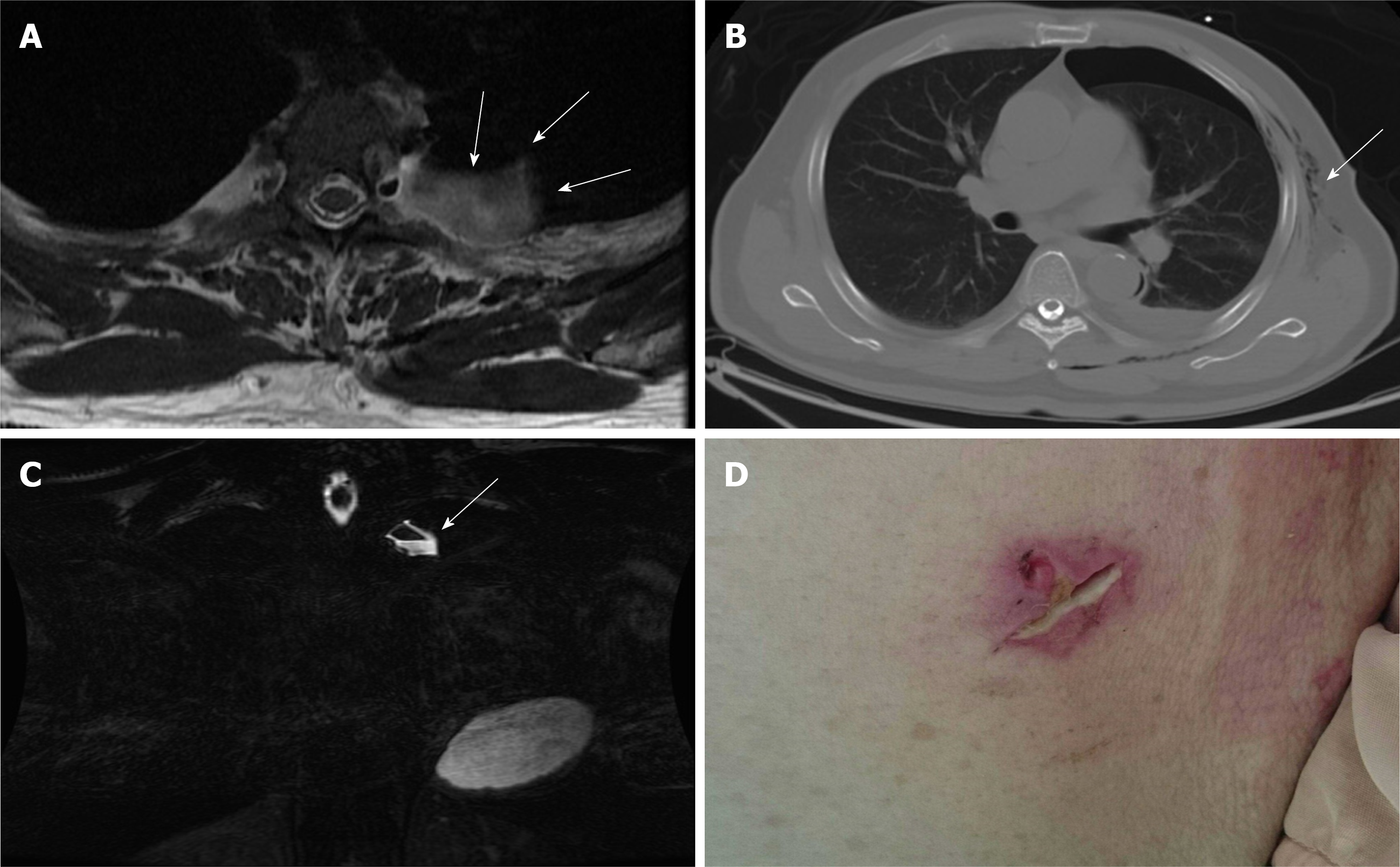
Figure 2 Pre-operative evaluations.
A-B: T2-weighted magnetic resonance imaging in axial view (A) and fast spin echo, fat-suppression coronal view (B) showing a cystic pouch laterally surrounding the spinal nerve root at left T3 level (arrow), which may be derived from the neural foramen of the L3 level. The air-fluid level was also demonstrated (arrow); C) Axial view of chest computed tomography showing pneumothorax and subcutaneous emphysema (arrow); D) Poorly healing previous thoracoscopic access wound.
- Citation: Chang CY, Hung CC, Liu JM, Chiu CD. Tension pneumocephalus following endoscopic resection of a mediastinal thoracic spinal tumor: A case report. World J Clin Cases 2022; 10(2): 725-732
- URL: https://www.wjgnet.com/2307-8960/full/v10/i2/725.htm
- DOI: https://dx.doi.org/10.12998/wjcc.v10.i2.725


