Copyright
©The Author(s) 2022.
World J Clin Cases. Jan 14, 2022; 10(2): 677-684
Published online Jan 14, 2022. doi: 10.12998/wjcc.v10.i2.677
Published online Jan 14, 2022. doi: 10.12998/wjcc.v10.i2.677
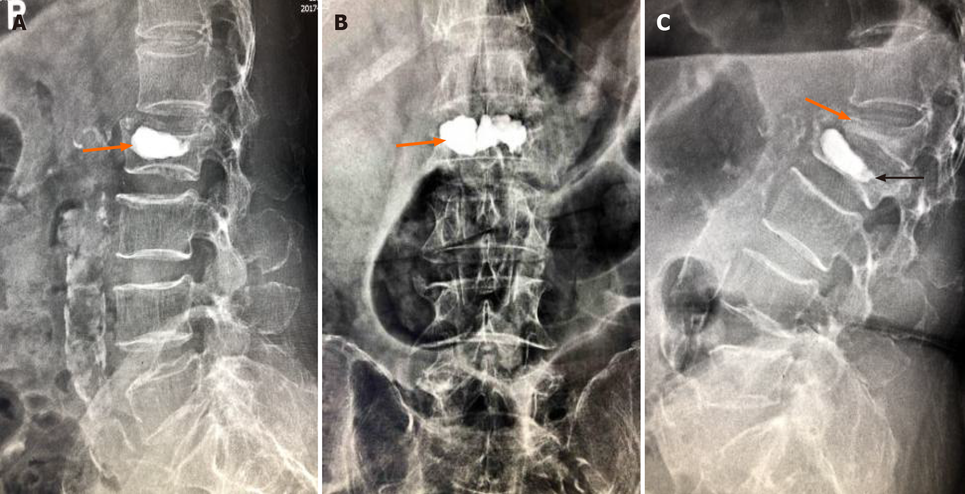
Figure 2 X-ray images.
A and B: Postoperative X-ray images showed that bone cement filled the cystic cavity of the L2 vertebral body with smooth edges, and there was insufficient bone cement dispersion outside the cystic cavity; C: Fifty days postoperatively, X-ray images showed L2 vertebral body collapse, slight forward bone cement displacement (indicated by the black arrow), L1 vertebral compression fracture, and severe collapse (indicated by the orange arrow).
- Citation: Li J, Liu Y, Peng L, Liu J, Cao ZD, He M. Intervertebral bridging ossification after kyphoplasty in a Parkinson’s patient with Kummell’s disease: A case report. World J Clin Cases 2022; 10(2): 677-684
- URL: https://www.wjgnet.com/2307-8960/full/v10/i2/677.htm
- DOI: https://dx.doi.org/10.12998/wjcc.v10.i2.677


