Copyright
©The Author(s) 2022.
World J Clin Cases. Jan 14, 2022; 10(2): 671-676
Published online Jan 14, 2022. doi: 10.12998/wjcc.v10.i2.671
Published online Jan 14, 2022. doi: 10.12998/wjcc.v10.i2.671
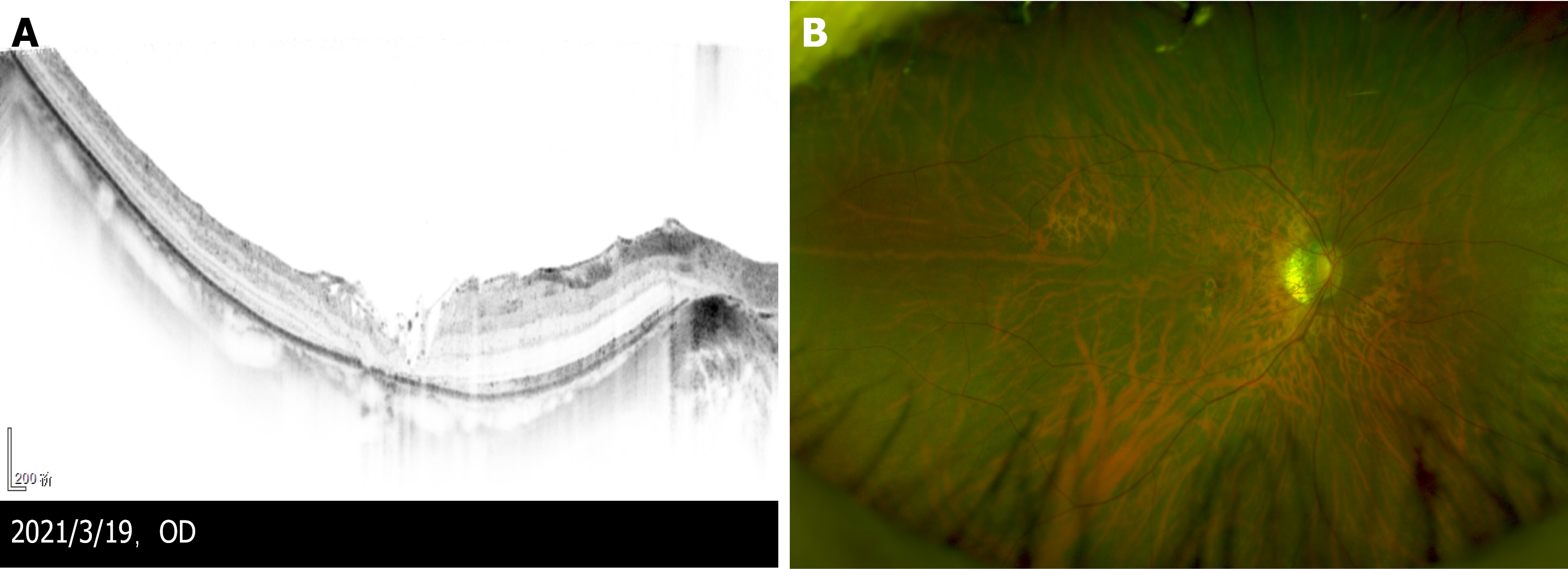
Figure 3 Manifestation of the eye 3 wk after the second vitrectomy.
A: Three weeks after the second vitrectomy, optical coherence tomography showed that the macular hole was closed, covered by flocculent internal limiting membrane. The split of each layer was significantly reduced, and photoreceptor cells were absent in the macular area; B: Optomap: Tessellated fundus only was observed.
- Citation: Ying HF, Wu SQ, Hu WP, Ni LY, Zhang ZL, Xu YG. Vitrectomy with residual internal limiting membrane covering and autologous blood for a secondary macular hole: A case report. World J Clin Cases 2022; 10(2): 671-676
- URL: https://www.wjgnet.com/2307-8960/full/v10/i2/671.htm
- DOI: https://dx.doi.org/10.12998/wjcc.v10.i2.671


