Copyright
©The Author(s) 2022.
World J Clin Cases. Jan 14, 2022; 10(2): 671-676
Published online Jan 14, 2022. doi: 10.12998/wjcc.v10.i2.671
Published online Jan 14, 2022. doi: 10.12998/wjcc.v10.i2.671
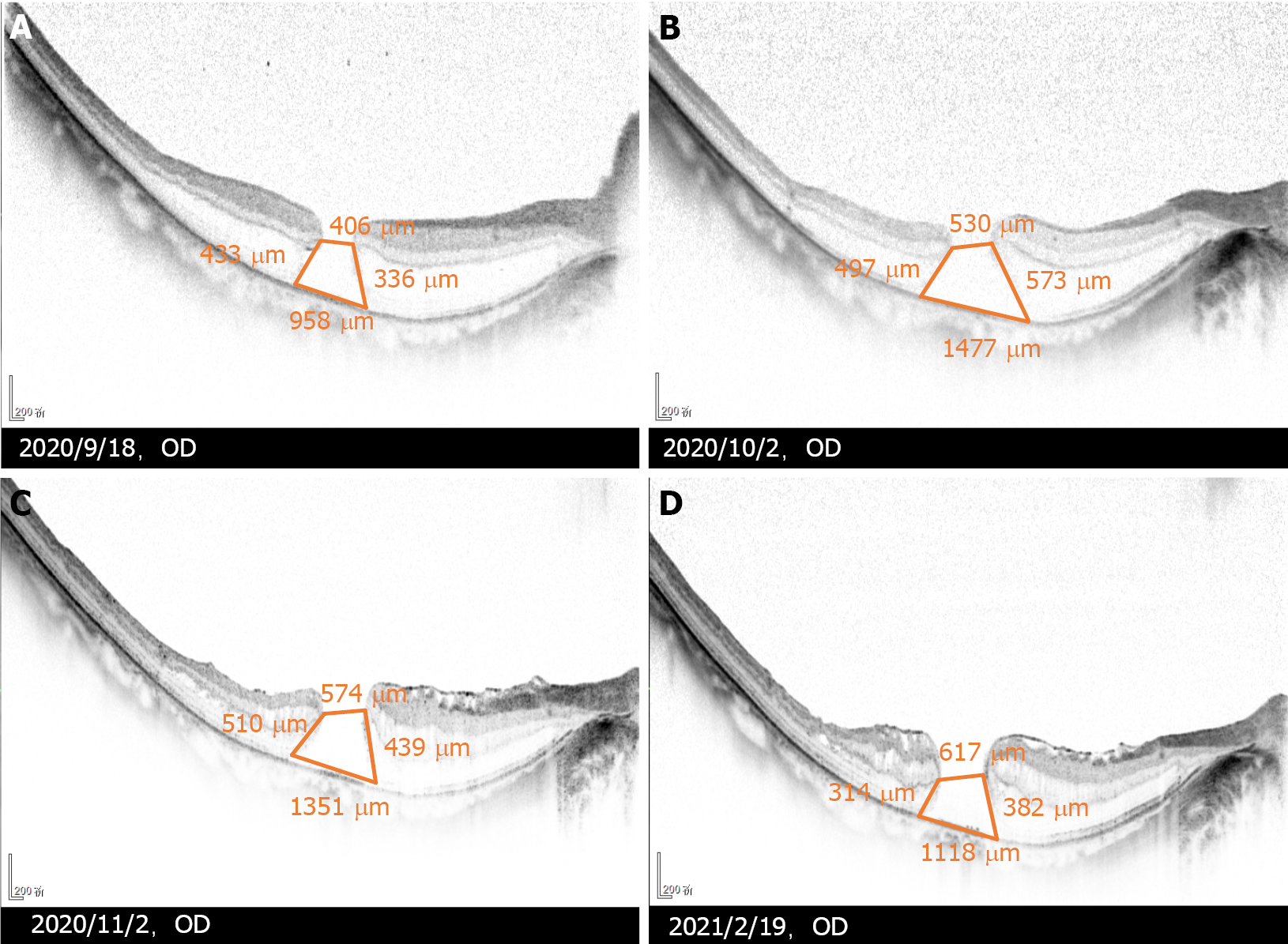
Figure 2 Changes of optical coherence tomography in the macular hole after initial vitrectomy.
A: One week postoperative; B: Three weeks postoperative; C: Two months postoperative; D: Five months postoperative. Five months after the initial vitrectomy, split range of the outer nuclear layer gradually reduced, a new split of the inner nuclear layer appeared, and the macular hole gradually expanded. The epiretinal membrane proliferated on the residual internal limiting membrane, wrinkled and caused the disordered shape of the nasal retinal nerve fibre layer.
- Citation: Ying HF, Wu SQ, Hu WP, Ni LY, Zhang ZL, Xu YG. Vitrectomy with residual internal limiting membrane covering and autologous blood for a secondary macular hole: A case report. World J Clin Cases 2022; 10(2): 671-676
- URL: https://www.wjgnet.com/2307-8960/full/v10/i2/671.htm
- DOI: https://dx.doi.org/10.12998/wjcc.v10.i2.671


