Copyright
©The Author(s) 2022.
World J Clin Cases. Jan 14, 2022; 10(2): 663-670
Published online Jan 14, 2022. doi: 10.12998/wjcc.v10.i2.663
Published online Jan 14, 2022. doi: 10.12998/wjcc.v10.i2.663
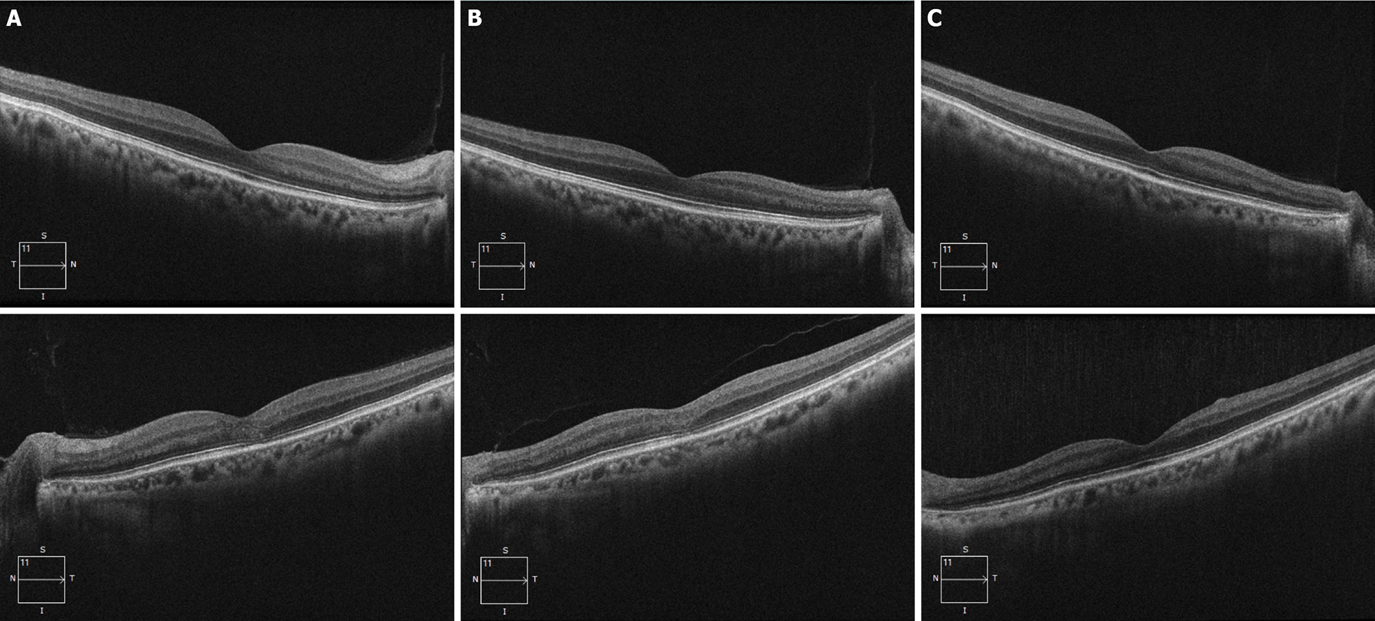
Figure 4 Outer nuclear layer high-definition optical coherence tomography.
A: At the initial visit, the outer nuclear layer under the fovea and the outer reflection bands representing the photoreceptor cells of the left eye were blurred (mainly the ellipsoid zone and the intersection area), and the damaged intersection area was mainly on the nasal side of the macula; B: Six months after drug withdrawal, compared with the first visit, the structure of the outer nuclear layer and ellipsoid zone of the macula had partially recovered; C: Eighteen months after drug withdrawl, the structure of the outer nuclear layer and the ellipsoid zone of the macula had recovered to normal appearance.
- Citation: Sheng WY, Wu SQ, Su LY, Zhu LW. Ethambutol-induced optic neuropathy with rare bilateral asymmetry onset: A case report. World J Clin Cases 2022; 10(2): 663-670
- URL: https://www.wjgnet.com/2307-8960/full/v10/i2/663.htm
- DOI: https://dx.doi.org/10.12998/wjcc.v10.i2.663


