Copyright
©The Author(s) 2022.
World J Clin Cases. Jan 14, 2022; 10(2): 663-670
Published online Jan 14, 2022. doi: 10.12998/wjcc.v10.i2.663
Published online Jan 14, 2022. doi: 10.12998/wjcc.v10.i2.663
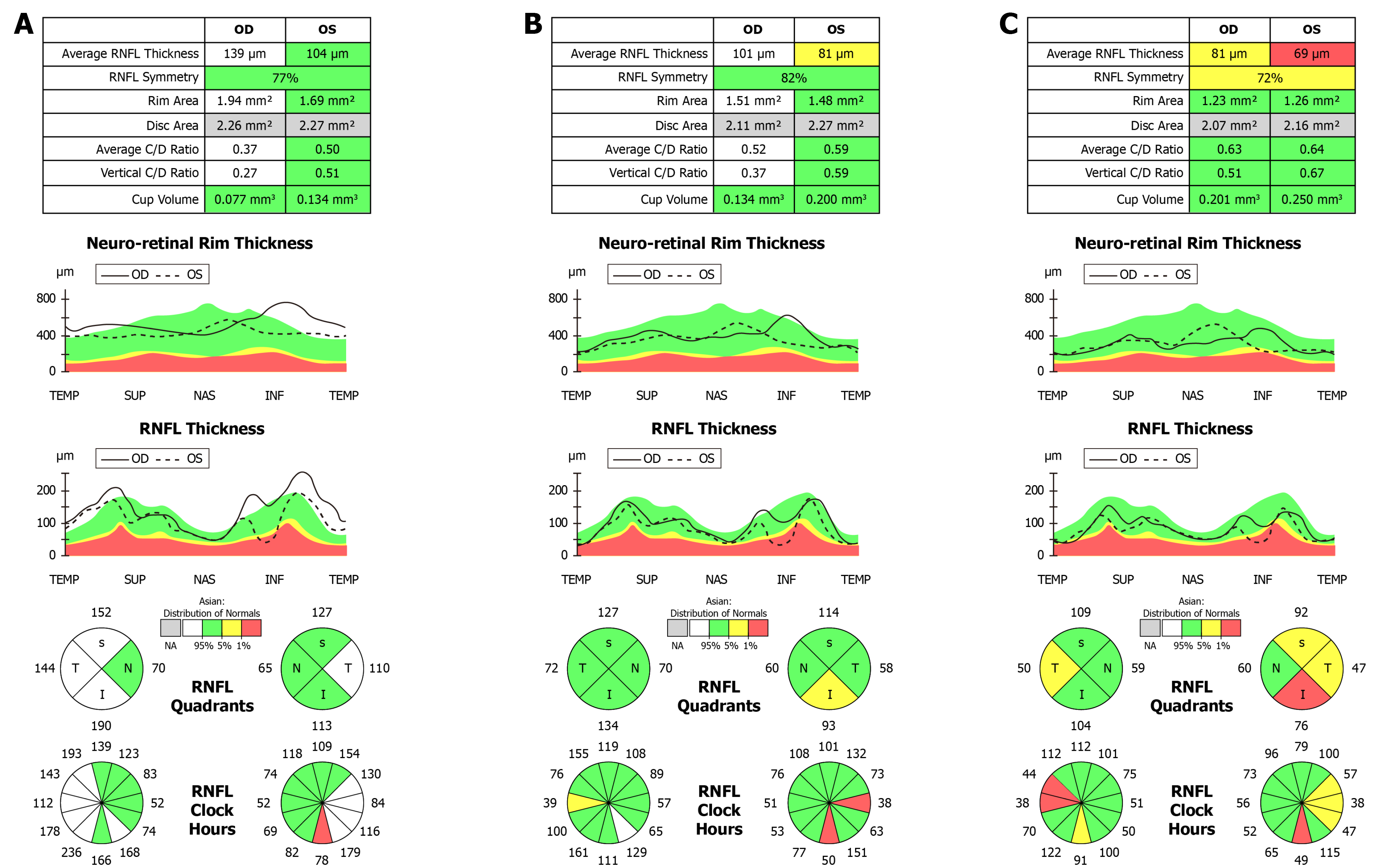
Figure 3 Retinal nerve fiber layer high-definition optical coherence tomography.
A: At the first visit, the retinal nerve fiber layer (RNFL) of the right eye had mild thickening in the superior, inferior and nasal regions; the RNFL of the left eye had mild thickening on the temporal side; B: Six months after drug withdrawal, the RNFL of both eyes became thinner compared with the first visit; the RNFL of the right eye was within the normal range; and the average and inferior thicknesses of the RNFL in the left eye were lower than normal; C: Eighteen months after stopping drug treatment, the RNFL of both eyes further decreased, and the temporal side of both eyes became thinner significantly.
- Citation: Sheng WY, Wu SQ, Su LY, Zhu LW. Ethambutol-induced optic neuropathy with rare bilateral asymmetry onset: A case report. World J Clin Cases 2022; 10(2): 663-670
- URL: https://www.wjgnet.com/2307-8960/full/v10/i2/663.htm
- DOI: https://dx.doi.org/10.12998/wjcc.v10.i2.663


