Copyright
©The Author(s) 2022.
World J Clin Cases. Jan 14, 2022; 10(2): 663-670
Published online Jan 14, 2022. doi: 10.12998/wjcc.v10.i2.663
Published online Jan 14, 2022. doi: 10.12998/wjcc.v10.i2.663
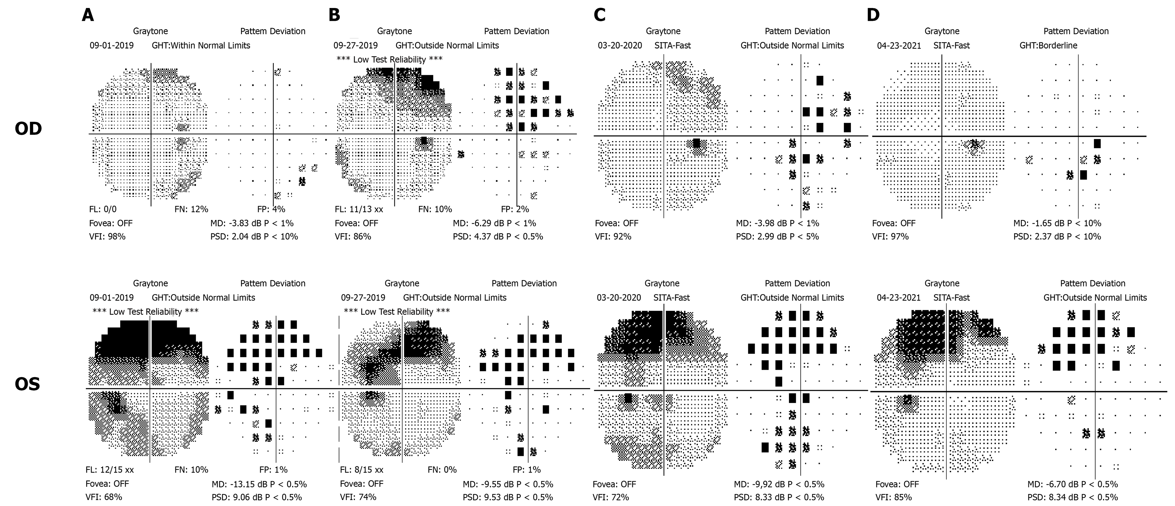
Figure 2 Computer perimetry results of four follow-up visits.
A: At the initial visit, there were suspicious dark spots in the inferonasal region of the right eye, and the visual field defect of the left eye manifested as concentric contraction; B: At the second visit, visual field defects had worsened in the right eye; C: At the third visit, the defect showed progression in the left eye, but improved in the right eye; D: At the fourth visit, the visual field in both eyes improved.
- Citation: Sheng WY, Wu SQ, Su LY, Zhu LW. Ethambutol-induced optic neuropathy with rare bilateral asymmetry onset: A case report. World J Clin Cases 2022; 10(2): 663-670
- URL: https://www.wjgnet.com/2307-8960/full/v10/i2/663.htm
- DOI: https://dx.doi.org/10.12998/wjcc.v10.i2.663


