Copyright
©The Author(s) 2022.
World J Clin Cases. Jan 14, 2022; 10(2): 631-642
Published online Jan 14, 2022. doi: 10.12998/wjcc.v10.i2.631
Published online Jan 14, 2022. doi: 10.12998/wjcc.v10.i2.631
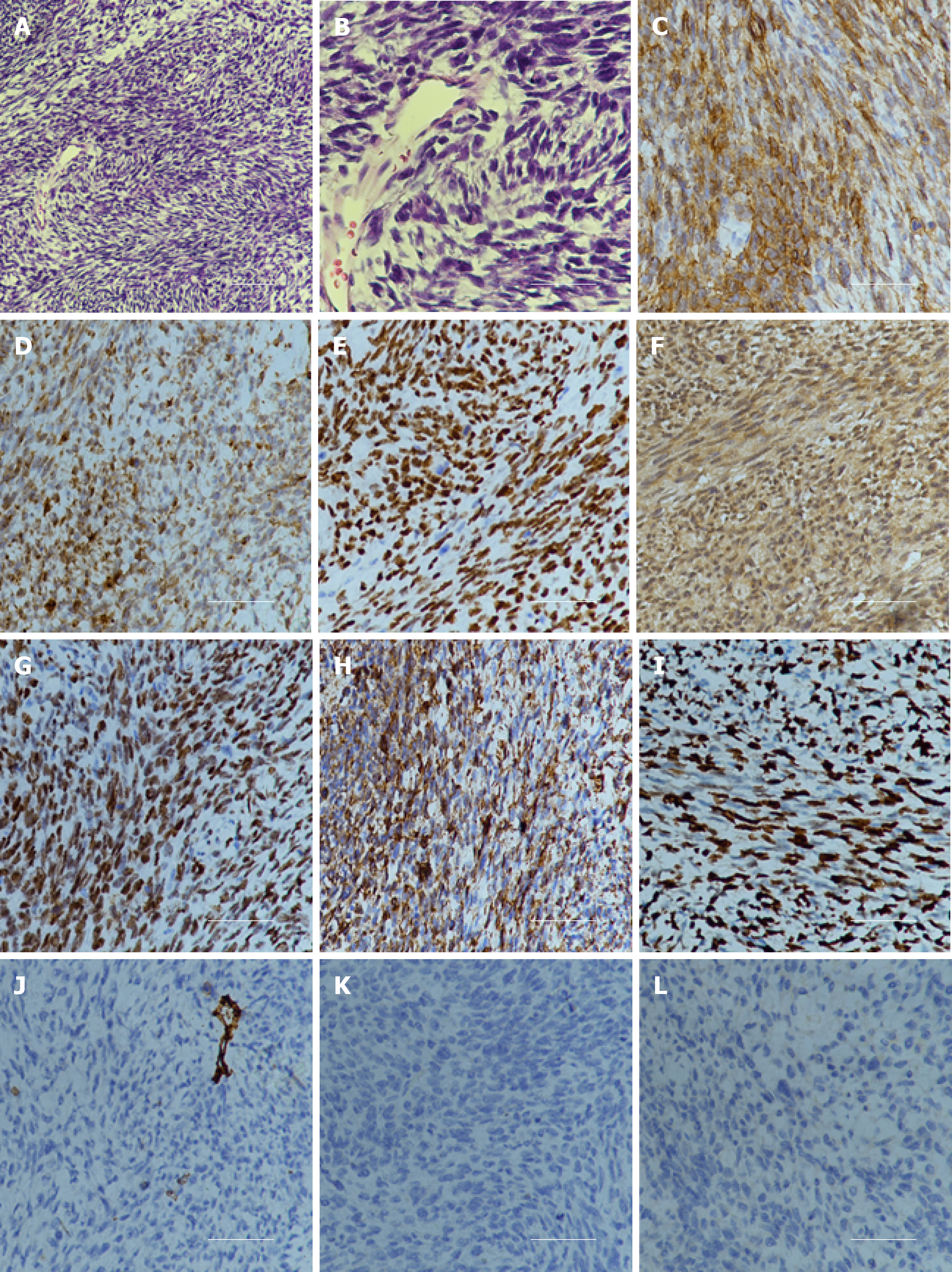
Figure 2 Hematoxylin and eosin staining and immunohistochemistry examination of the specimen.
A: Hematoxylin and eosin staining showed that a large number of spindle or oval cells were diffusely distributed, with deep staining of a null, “staghorn” vascular pattern, hypercellularity and increased mitotic activity were observed in the tumor (> 4 mitosis/10 high-power fields). Hematoxylin and eosin, 100 ×; B: Hematoxylin and eosin staining, 400 ×; C: Positive for CD99, 200 ×; D: Positive for Bcl-2, 200 ×; E: Positive for TP53, 200 ×; F: Positive for IDH1, 200 ×; G: Positive for TLE-1, 200 ×; H: Positive for vimentin, 200 ×; I: High Ki-67 proliferation index: 80%, 200 ×; J: Negative for CD34, 200 ×; K: Negative for STAT6, 200 ×; L: Negative for S100, 200 ×.
- Citation: Zhang DY, Su L, Wang YW. Malignant solitary fibrous tumor in the central nervous system treated with surgery, radiotherapy and anlotinib: A case report. World J Clin Cases 2022; 10(2): 631-642
- URL: https://www.wjgnet.com/2307-8960/full/v10/i2/631.htm
- DOI: https://dx.doi.org/10.12998/wjcc.v10.i2.631


