Copyright
©The Author(s) 2022.
World J Clin Cases. Jan 14, 2022; 10(2): 477-484
Published online Jan 14, 2022. doi: 10.12998/wjcc.v10.i2.477
Published online Jan 14, 2022. doi: 10.12998/wjcc.v10.i2.477
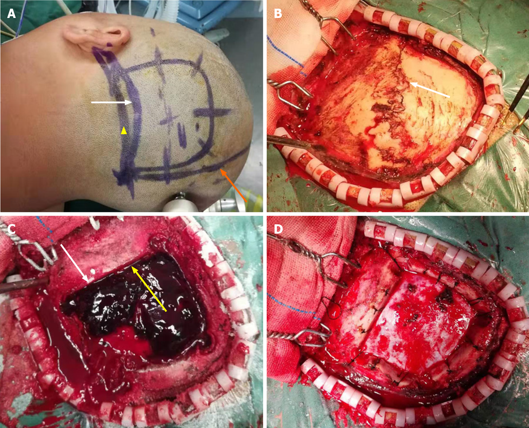
Figure 3 Modified surgical method of supra- and infratentorial epidural hematoma.
A: The white arrow points to the surface projection of the transverse sinus. The skin flap was close to the midline medially, reaching the lateral edge of the hematoma laterally, up to the upper edge of the hematoma superiorly, and about 1 cm below the transverse sinus inferiorly (yellow triangle). The lower edge of the bone flap was located at the upper edge of the transverse sinus. The orange arrow indicates the midline of the head; B: After elevating the skin flap, a linear fracture of the occipital bone was found (white arrow); C: Subsequently the bone flap was opened to expose the supra- and infratentorial epidural hematoma (SIEDH). The white arrow shows the lower edge of the bone window across the superior edge of the transverse sinus. The yellow arrow demonstrated that the SIEDH can be cleared from above to below the transverse sinus; D: After the SIEDH was completely removed, the dura was tightly suspended on the periosteum.
- Citation: Li RC, Guo SW, Liang C. Modified surgical method of supra- and infratentorial epidural hematoma and the related anatomical study of the squamous part of the occipital bone. World J Clin Cases 2022; 10(2): 477-484
- URL: https://www.wjgnet.com/2307-8960/full/v10/i2/477.htm
- DOI: https://dx.doi.org/10.12998/wjcc.v10.i2.477


