Copyright
©The Author(s) 2022.
World J Clin Cases. Jan 14, 2022; 10(2): 477-484
Published online Jan 14, 2022. doi: 10.12998/wjcc.v10.i2.477
Published online Jan 14, 2022. doi: 10.12998/wjcc.v10.i2.477
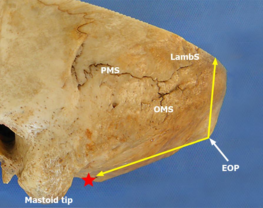
Figure 1 Schematic representation of the angle of the squamous part of the occipital bone on the outer surface of the occipital bone.
The vertex of the angle of the squamous part of the occipital bone was located at the external occipital protuberance (EOP), while the two edges of the angle were demonstrated by two yellow arrow lines directed from the EOP to the lambdoid suture and the posterior edge of the foramen magnum (red star) in the median sagittal plane, respectively. OMS: Occipitomastoid suture; PMS: Parietomastoid suture; LambS: Lambdoid suture; EOP: External occipital protuberance.
- Citation: Li RC, Guo SW, Liang C. Modified surgical method of supra- and infratentorial epidural hematoma and the related anatomical study of the squamous part of the occipital bone. World J Clin Cases 2022; 10(2): 477-484
- URL: https://www.wjgnet.com/2307-8960/full/v10/i2/477.htm
- DOI: https://dx.doi.org/10.12998/wjcc.v10.i2.477


