Copyright
©The Author(s) 2022.
World J Clin Cases. Jul 6, 2022; 10(19): 6750-6758
Published online Jul 6, 2022. doi: 10.12998/wjcc.v10.i19.6750
Published online Jul 6, 2022. doi: 10.12998/wjcc.v10.i19.6750
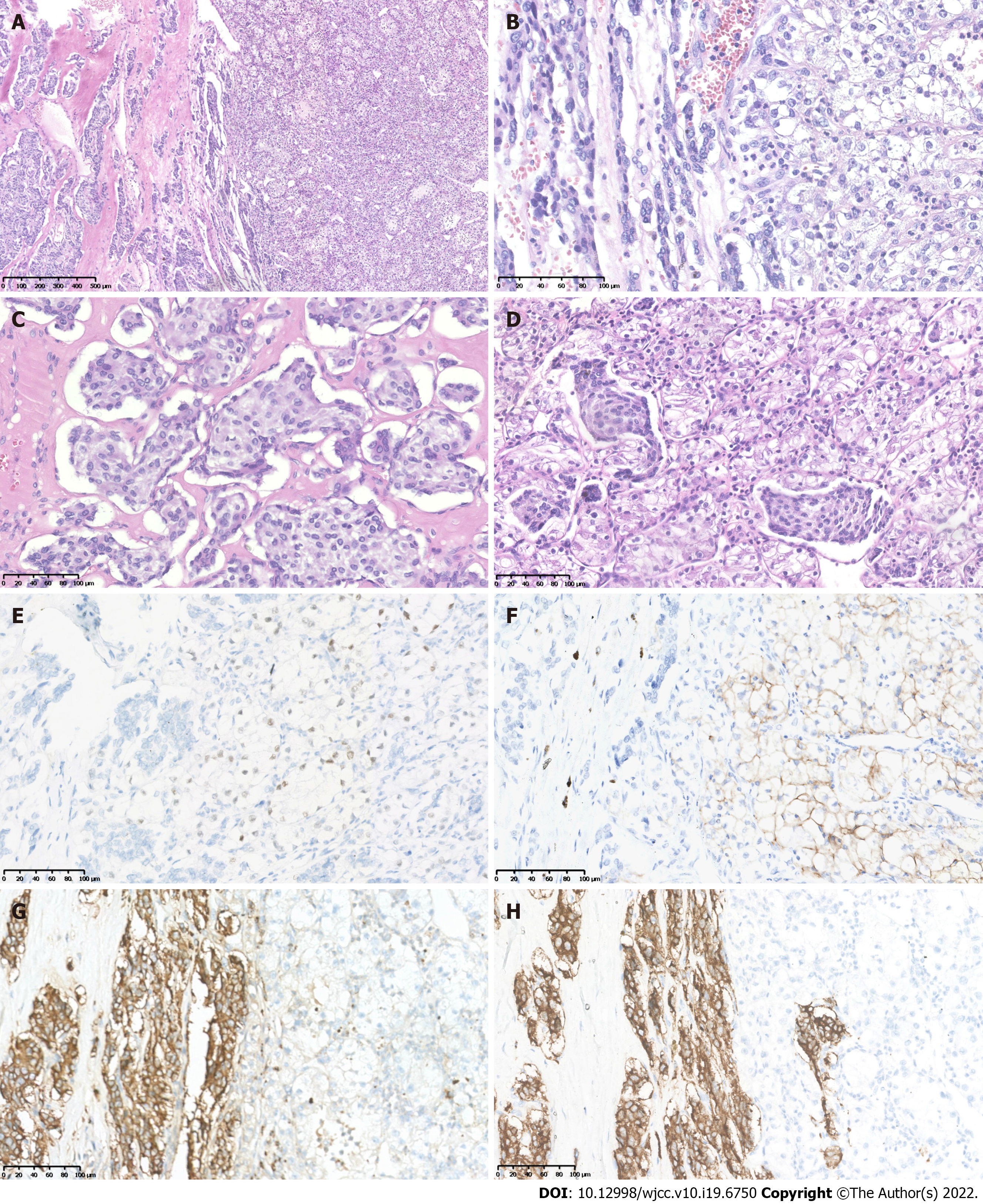
Figure 3 Histopathological and immunohistochemiscal figures showed the left retroperitoneal tumor mass contained two components.
A: One component was typical pheochromocytoma (PCC) with tumor cells arranged in organoid nests and cords; B: The other component exhibited clear cell renal cell carcinoma (CCRCC) morphology; C: The PCC nests at high magnification; D: The PCC nests mixed with CCRCC at their intersection; E: Immunohistochemistry (IHC) showed PAX8 was positive in the CCRCC tumor cells; F: IHC showed CAIX was positive in the CCRCC tumor cells; G: PCC tumor cells showed positive for CgA; H: PCC tumor cells showed positive for Syn.
- Citation: Wen HY, Hou J, Zeng H, Zhou Q, Chen N. Tumor-to-tumor metastasis of clear cell renal cell carcinoma to contralateral synchronous pheochromocytoma: A case report. World J Clin Cases 2022; 10(19): 6750-6758
- URL: https://www.wjgnet.com/2307-8960/full/v10/i19/6750.htm
- DOI: https://dx.doi.org/10.12998/wjcc.v10.i19.6750


