Copyright
©The Author(s) 2022.
World J Clin Cases. Jul 6, 2022; 10(19): 6710-6715
Published online Jul 6, 2022. doi: 10.12998/wjcc.v10.i19.6710
Published online Jul 6, 2022. doi: 10.12998/wjcc.v10.i19.6710
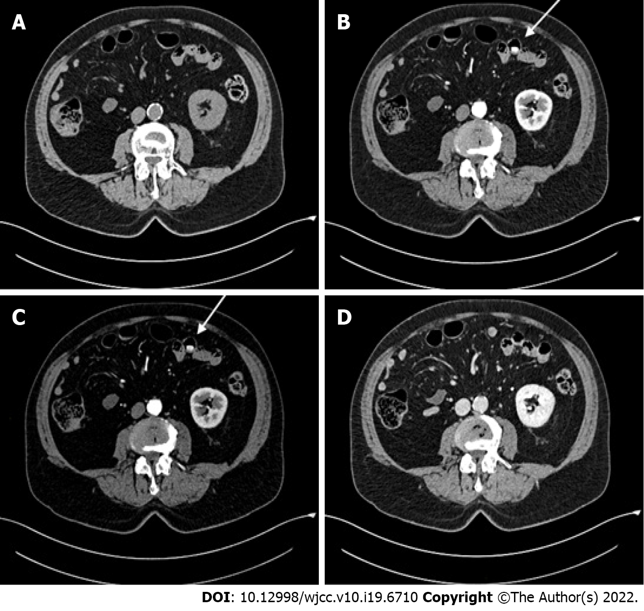
Figure 1 Abdominal contrast-enhanced computed tomography scan.
A: Pre-contrast phase; B: Arterial phase; C: Venous phase; D: Delayed-contrast phase. A hyperdense jejunal image (white arrow) was observed in the arterial and venous phases.
- Citation: Frosio F, Rausa E, Marra P, Boutron-Ruault MC, Lucianetti A. Delayed-release oral mesalamine tablet mimicking a small jejunal gastrointestinal stromal tumor: A case report. World J Clin Cases 2022; 10(19): 6710-6715
- URL: https://www.wjgnet.com/2307-8960/full/v10/i19/6710.htm
- DOI: https://dx.doi.org/10.12998/wjcc.v10.i19.6710


