Copyright
©The Author(s) 2022.
World J Clin Cases. Jul 6, 2022; 10(19): 6626-6635
Published online Jul 6, 2022. doi: 10.12998/wjcc.v10.i19.6626
Published online Jul 6, 2022. doi: 10.12998/wjcc.v10.i19.6626
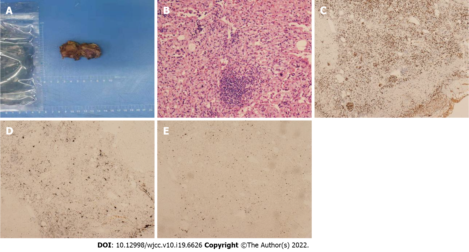
Figure 5 Intrahepatic extramedullary hematopoiesis in the same patient shown in figure 3.
A: Photograph of the specimen showed the lobular and solid nature of the resected hepatic mass (segment V/VIII), without areas of necrosis and hemorrhage; B: On the photomicrograph (hematoxylin and eosin staining; × 200), granulocytes, megakaryocytes, adipocytes and erythrocytes were distributed within the surgical specimen; C-E: Immunohistochemical staining using CD235 (C), CD61 (D) and MPO (E) markers (× 40) revealed that the cells were positive (brown color) for these markers, respectively.
- Citation: Luo M, Chen JW, Xie CM. Magnetic resonance imaging features of intrahepatic extramedullary hematopoiesis: Three case reports. World J Clin Cases 2022; 10(19): 6626-6635
- URL: https://www.wjgnet.com/2307-8960/full/v10/i19/6626.htm
- DOI: https://dx.doi.org/10.12998/wjcc.v10.i19.6626


