Copyright
©The Author(s) 2022.
World J Clin Cases. Jul 6, 2022; 10(19): 6626-6635
Published online Jul 6, 2022. doi: 10.12998/wjcc.v10.i19.6626
Published online Jul 6, 2022. doi: 10.12998/wjcc.v10.i19.6626
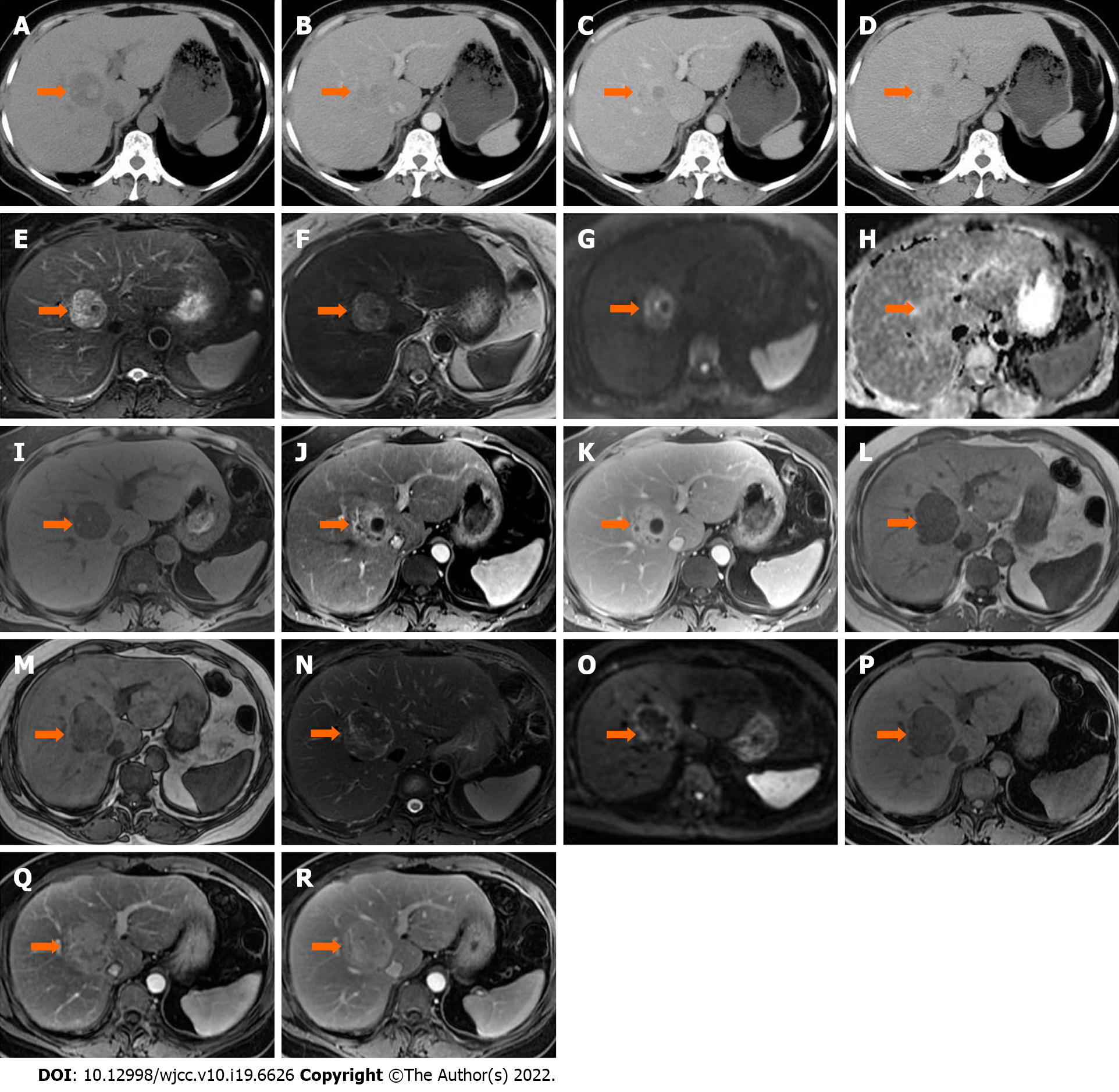
Figure 1 An intrahepatic mass (arrow) in segment VIII was found in a 50-year-old female.
A: On unenhanced computed tomography, the lesion was heterogeneously hypodense, with hyperdense foci in the central area; B: The lesion was heterogeneously hyperdense in the arterial phase; C and D: Progressive enhancement in the portal venous (C) and delayed phases (D); E-K: On magnetic resonance imaging, the lesion was heterogeneously hyperintense on T2 weighted image (WI), T2WI-fat saturation (FS) (E and F) and diffusion weighted imaging (DWI) (G), isointense on the apparent diffusion coefficient map (H), hypointense on T1WI-FS (I), avid enhancement in the arterial phase (J) and persistent enhancement in the delayed phase (K); L-R: Corresponding follow-up images five months later showed that the lesion size had increased. Signal drop was seen on the in-phase (L) compared with the out-phase (M) image. The lesion was heterogeneously hypointense on T2WI-FS (N) and DWI (O), homogeneously hypointense on T1WI-FS (P), and showed hypervascular enhancement with delayed enhancement in the arterial (Q) and delayed phase (R), respectively. The surgical pathologic diagnosis was intrahepatic extramedullary hematopoiesis.
- Citation: Luo M, Chen JW, Xie CM. Magnetic resonance imaging features of intrahepatic extramedullary hematopoiesis: Three case reports. World J Clin Cases 2022; 10(19): 6626-6635
- URL: https://www.wjgnet.com/2307-8960/full/v10/i19/6626.htm
- DOI: https://dx.doi.org/10.12998/wjcc.v10.i19.6626


