Copyright
©The Author(s) 2022.
World J Clin Cases. Jul 6, 2022; 10(19): 6609-6616
Published online Jul 6, 2022. doi: 10.12998/wjcc.v10.i19.6609
Published online Jul 6, 2022. doi: 10.12998/wjcc.v10.i19.6609
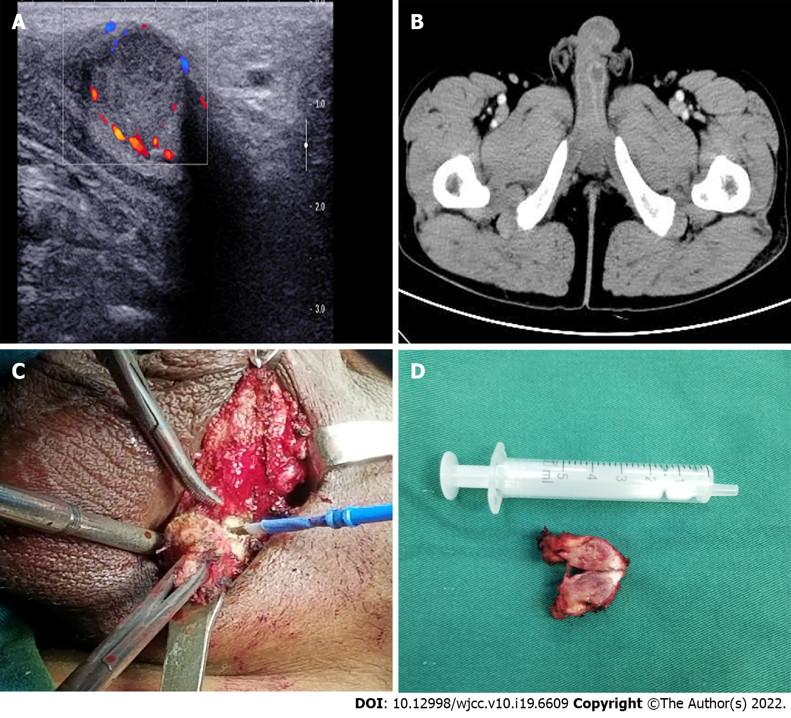
Figure 1 Imaging and macroscopic examination of the secondary penile tumour.
A: Ultrasonography showed a hyper-echoic mass on the root of the left penis; B: Abdominal contrast-enhanced computed tomography indicated a nodular mass measuring 1.5 cm with mild enhancement; C: A rigid nodule with distinct margins found in the left penis during the operation; D: The resected specimen measured 1.5 cm in diameter.
- Citation: Sun JJ, Zhang SY, Tian JJ, Jin BY. Penile metastasis from rectal carcinoma: A case report. World J Clin Cases 2022; 10(19): 6609-6616
- URL: https://www.wjgnet.com/2307-8960/full/v10/i19/6609.htm
- DOI: https://dx.doi.org/10.12998/wjcc.v10.i19.6609


