Copyright
©The Author(s) 2022.
World J Clin Cases. Jul 6, 2022; 10(19): 6595-6601
Published online Jul 6, 2022. doi: 10.12998/wjcc.v10.i19.6595
Published online Jul 6, 2022. doi: 10.12998/wjcc.v10.i19.6595
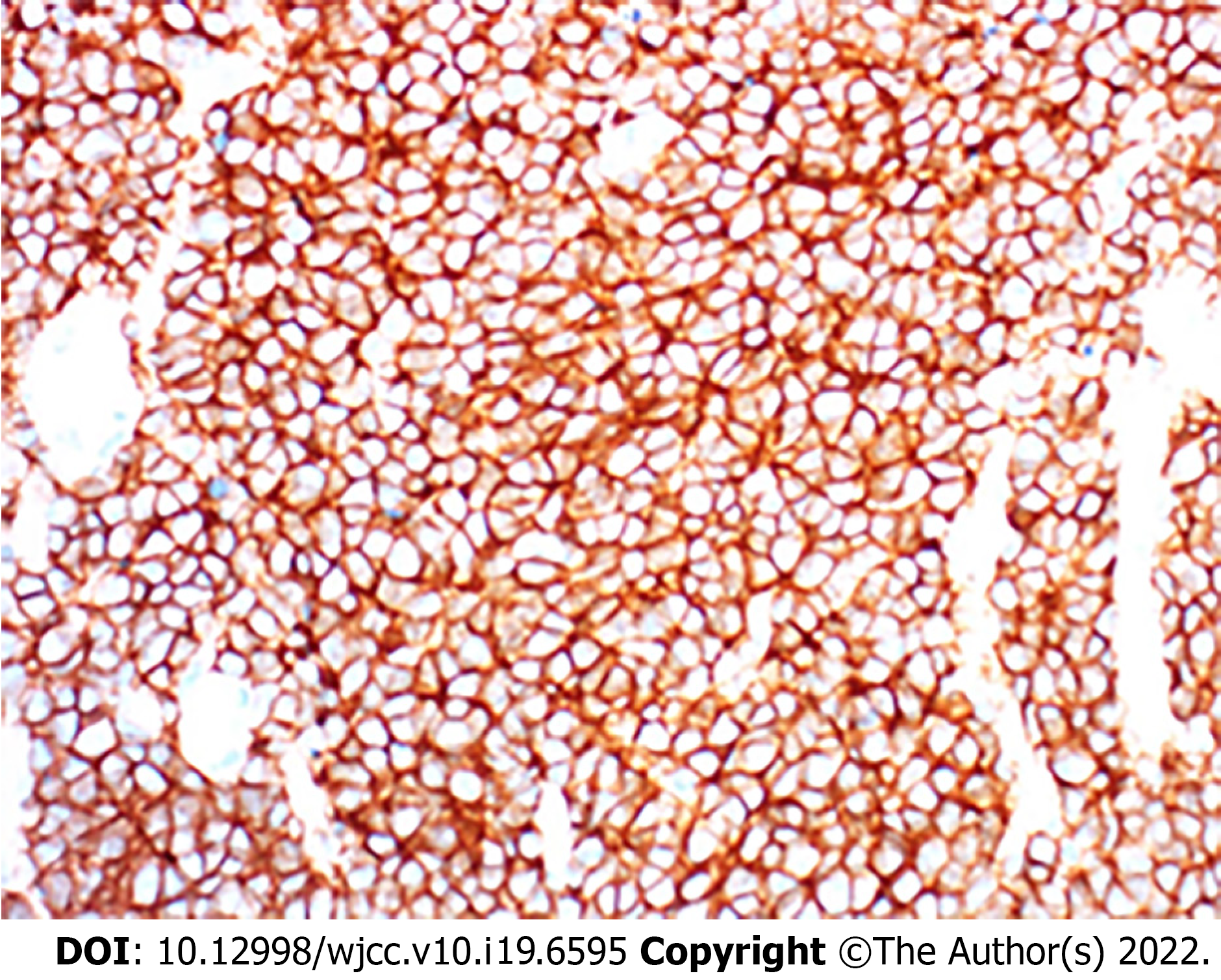
Figure 4 Histologic specimen (immunohistochemical stain, immunohistochemistry; original magnification, × 400) confirmed a diagnosis of the extraskeletal Ewing sarcoma, with the small round blue-stained cells observed.
Immunochemical histological findings: NSE(+), NF(-), INI-1(+++), TTF-1(-), CK20(-), TDT(-), Wilms tumor(-), Bcl-2(+++), Ki-67(+), CD34(-), CD99(+++), Vimentin(+++), Syn(+).
- Citation: Chen ZH, Guo HQ, Chen JJ, Zhang Y, Zhao L. Imaging-based diagnosis for extraskeletal Ewing sarcoma in pediatrics: A case report . World J Clin Cases 2022; 10(19): 6595-6601
- URL: https://www.wjgnet.com/2307-8960/full/v10/i19/6595.htm
- DOI: https://dx.doi.org/10.12998/wjcc.v10.i19.6595


