Copyright
©The Author(s) 2022.
World J Clin Cases. Jul 6, 2022; 10(19): 6563-6570
Published online Jul 6, 2022. doi: 10.12998/wjcc.v10.i19.6563
Published online Jul 6, 2022. doi: 10.12998/wjcc.v10.i19.6563
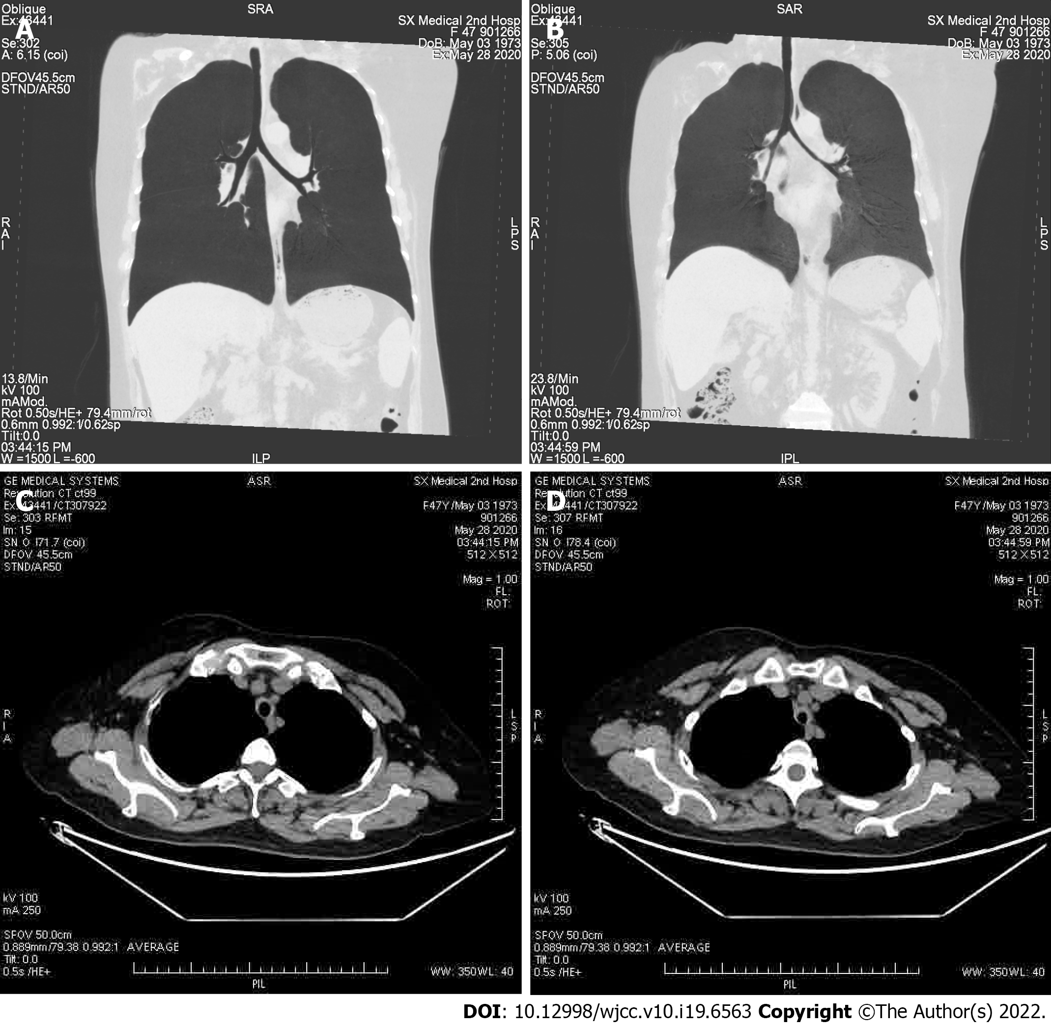
Figure 3 Computed tomography analysis.
A and B: The diameters of the trachea and bilateral bronchi show narrowing in the expiratory phase (B) compared to the inspiratory phase (A); C and D: The axial computed tomography image of mediastinal field for inspiration (C) and expiration (D).
- Citation: Chen JY, Li XY, Zong C. Relapsing polychondritis with isolated tracheobronchial involvement complicated with Sjogren's syndrome: A case report. World J Clin Cases 2022; 10(19): 6563-6570
- URL: https://www.wjgnet.com/2307-8960/full/v10/i19/6563.htm
- DOI: https://dx.doi.org/10.12998/wjcc.v10.i19.6563


