Copyright
©The Author(s) 2022.
World J Clin Cases. Jul 6, 2022; 10(19): 6536-6542
Published online Jul 6, 2022. doi: 10.12998/wjcc.v10.i19.6536
Published online Jul 6, 2022. doi: 10.12998/wjcc.v10.i19.6536
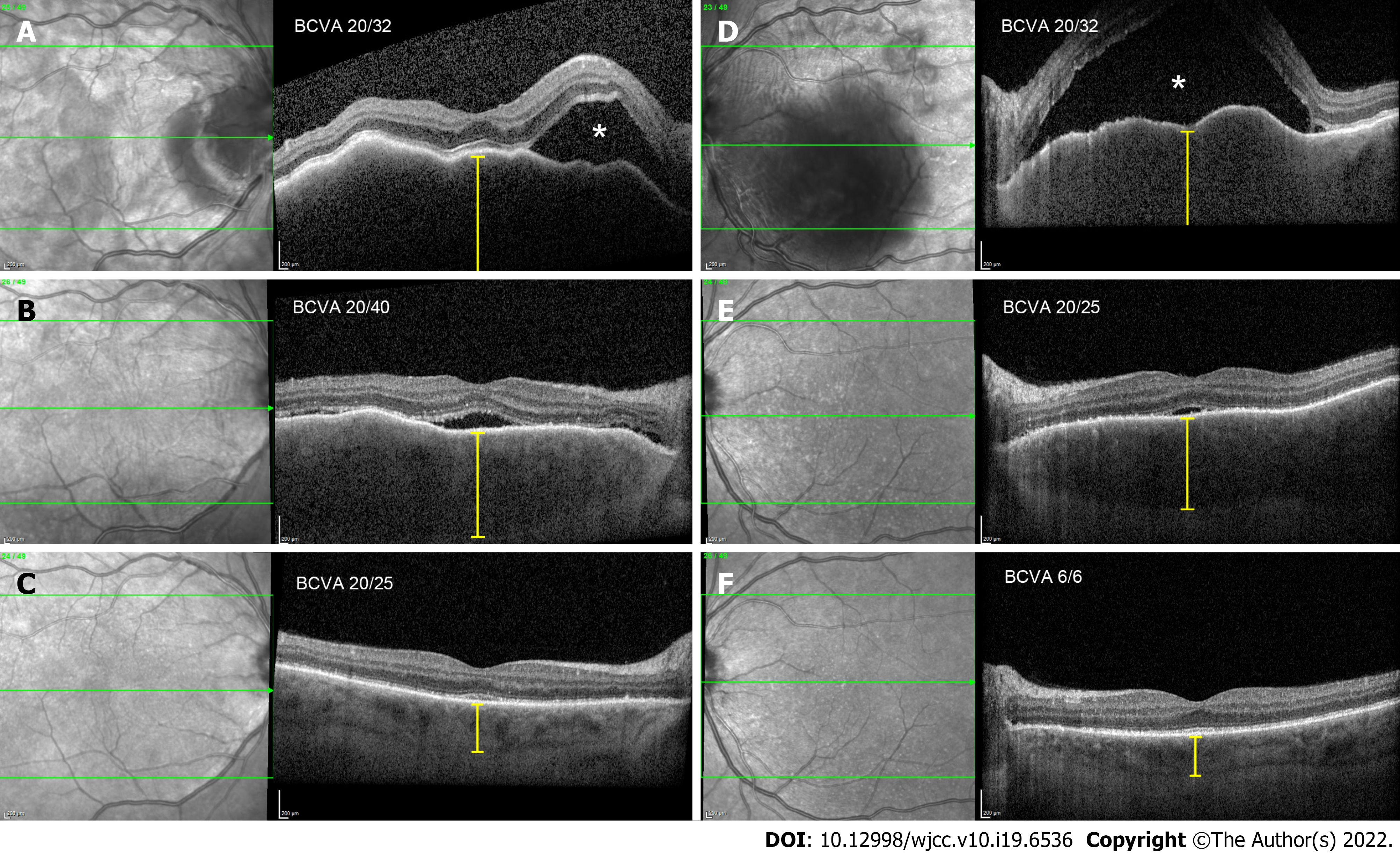
Figure 1 Optical coherence tomography morphological changes at presentation, 2 wk later, and 1 mo later.
A (RE) and D (LE): Bullous exudative retinal detachment (white asterisk), chorioretinal folds, and thickened choroid (yellow caliper), at presentation; B (RE) and E (LE): Substantial resorption of subretinal fluid and partial resolution of chorioretinal folds and choroidal thickness, after 2 wk; C (RE) and F (LE): Complete resolution of subretinal fluid and chorioretinal folds, normal choroidal thickness, after 1 mo.
- Citation: Kiraly P, Groznik AL, Valentinčič NV, Mekjavić PJ, Urbančič M, Ocvirk J, Mesti T. Choroidal thickening with serous retinal detachment in BRAF/MEK inhibitor-induced uveitis: A case report. World J Clin Cases 2022; 10(19): 6536-6542
- URL: https://www.wjgnet.com/2307-8960/full/v10/i19/6536.htm
- DOI: https://dx.doi.org/10.12998/wjcc.v10.i19.6536


