Copyright
©The Author(s) 2022.
World J Clin Cases. Jul 6, 2022; 10(19): 6428-6436
Published online Jul 6, 2022. doi: 10.12998/wjcc.v10.i19.6428
Published online Jul 6, 2022. doi: 10.12998/wjcc.v10.i19.6428
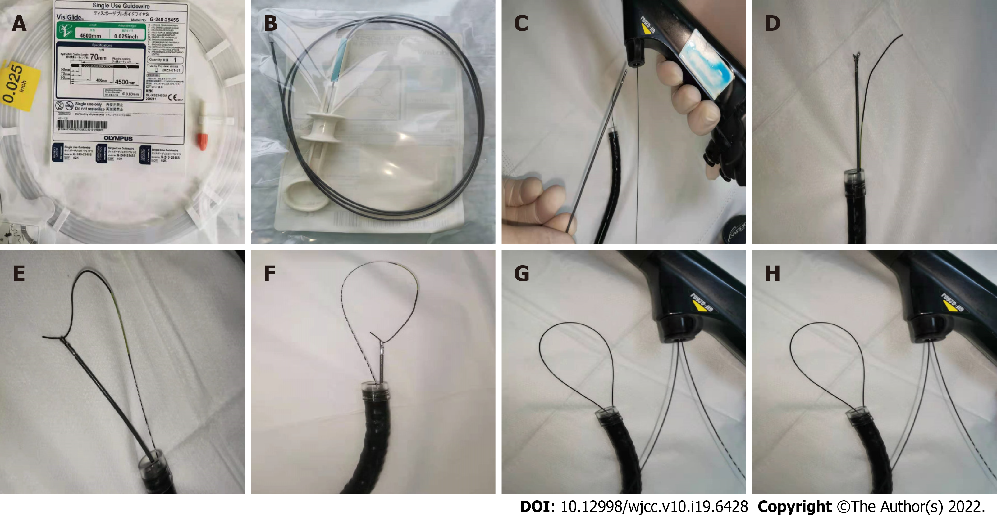
Figure 1 Generation of the wire loop snares.
A: A 0.025-inch diameter guidewire (Single-Use Guidewire; Olympus, Tokyo, Japan); B: An ultrafine endoscopic biopsy forcep (FB-231K; Olympus); C: The guidewire is pushed into the biopsy channel, and biopsy forceps is inserted into the biopsy channel afterward; D: The guidewire and biopsy forceps are passed through the endoscopic channel, and the front end of the gastroscope is covered by a transparent cap; E: The guidewire is passed through the lateral orifice at the head end of the biopsy forceps; F: The biopsy forceps are closed to clamp the guidewire; G: The biopsy forceps are pulled back until the guidewire is pulled out of the biopsy channel. During the procedure, the guidewire is delivered into the biopsy channel synchronously to keep an “O” shape of the loop snare extending from the biopsy channel; H: A guidewire loop snare with a variable diameter.
- Citation: Xu W, Liu XB, Li SB, Deng WP, Tong Q. Self-made wire loop snare successfully treats gastric persimmon stone under endoscopy. World J Clin Cases 2022; 10(19): 6428-6436
- URL: https://www.wjgnet.com/2307-8960/full/v10/i19/6428.htm
- DOI: https://dx.doi.org/10.12998/wjcc.v10.i19.6428


