Copyright
©The Author(s) 2022.
World J Clin Cases. Jun 26, 2022; 10(18): 6289-6297
Published online Jun 26, 2022. doi: 10.12998/wjcc.v10.i18.6289
Published online Jun 26, 2022. doi: 10.12998/wjcc.v10.i18.6289
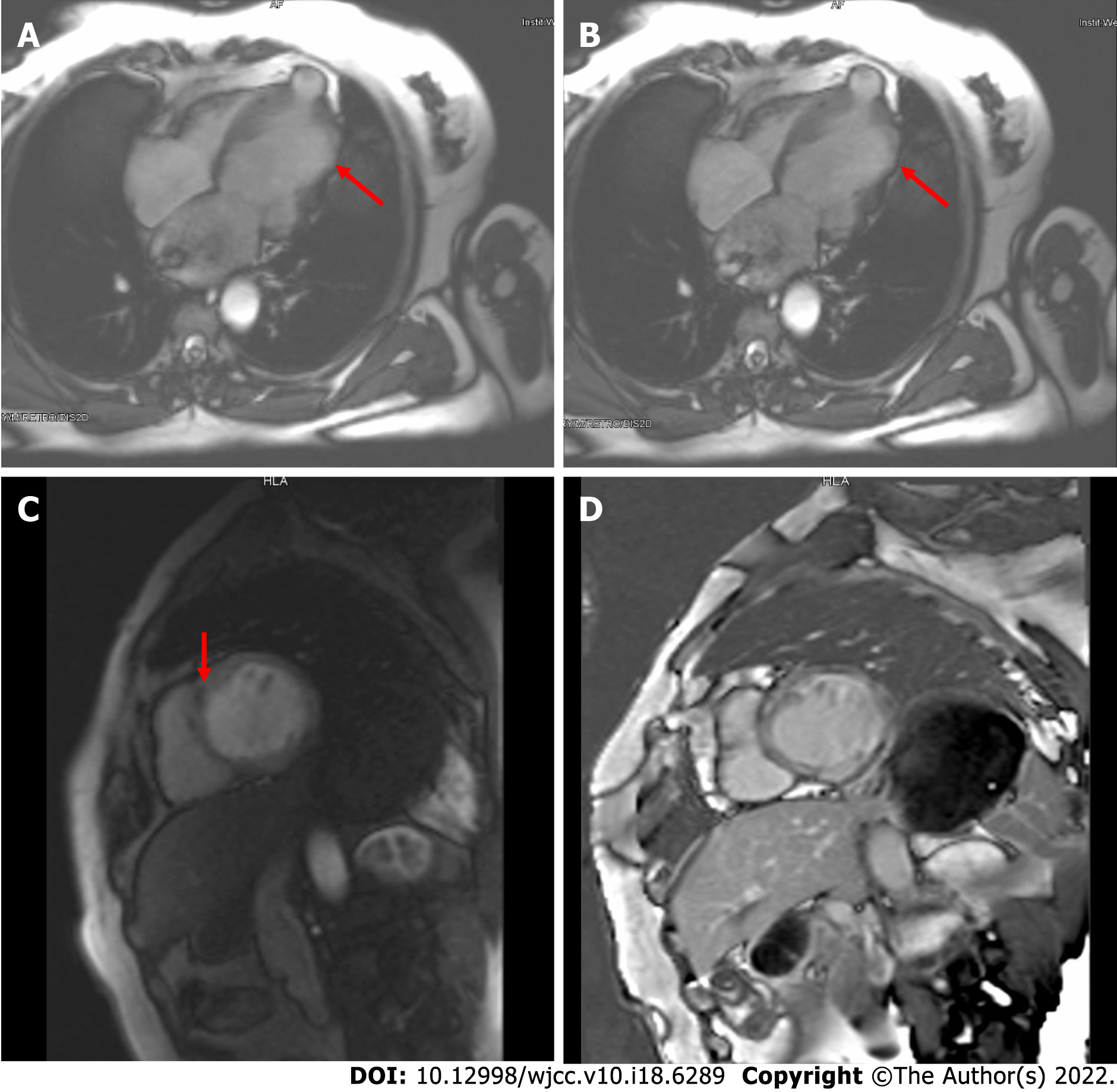
Figure 6 Cardiac magnetic resonance imaging results of the patient under this study.
A and B: Four-chamber cine images at end-diastole (A) and end-systole (B) showed left ventricular dilatation, abnormal activity of the ventricular wall (arrows indicate severely diminished contractility), and outpouching at the left ventricular apex; C: Short-axis first-pass perfusion showed a low signal at the midwall of the septum myocardium; D: Short-axis delayed enhancement imaging demonstrated diffuse stripes of hyperenhancement in the midwall of the left ventricular septum and free wall myocardium.
- Citation: Zhang X, Ye RY, Chen XP. Dilated left ventricle with multiple outpouchings — a severe congenital ventricular diverticulum or left-dominant arrhythmogenic cardiomyopathy: A case report. World J Clin Cases 2022; 10(18): 6289-6297
- URL: https://www.wjgnet.com/2307-8960/full/v10/i18/6289.htm
- DOI: https://dx.doi.org/10.12998/wjcc.v10.i18.6289


