Copyright
©The Author(s) 2022.
World J Clin Cases. Jun 26, 2022; 10(18): 6289-6297
Published online Jun 26, 2022. doi: 10.12998/wjcc.v10.i18.6289
Published online Jun 26, 2022. doi: 10.12998/wjcc.v10.i18.6289
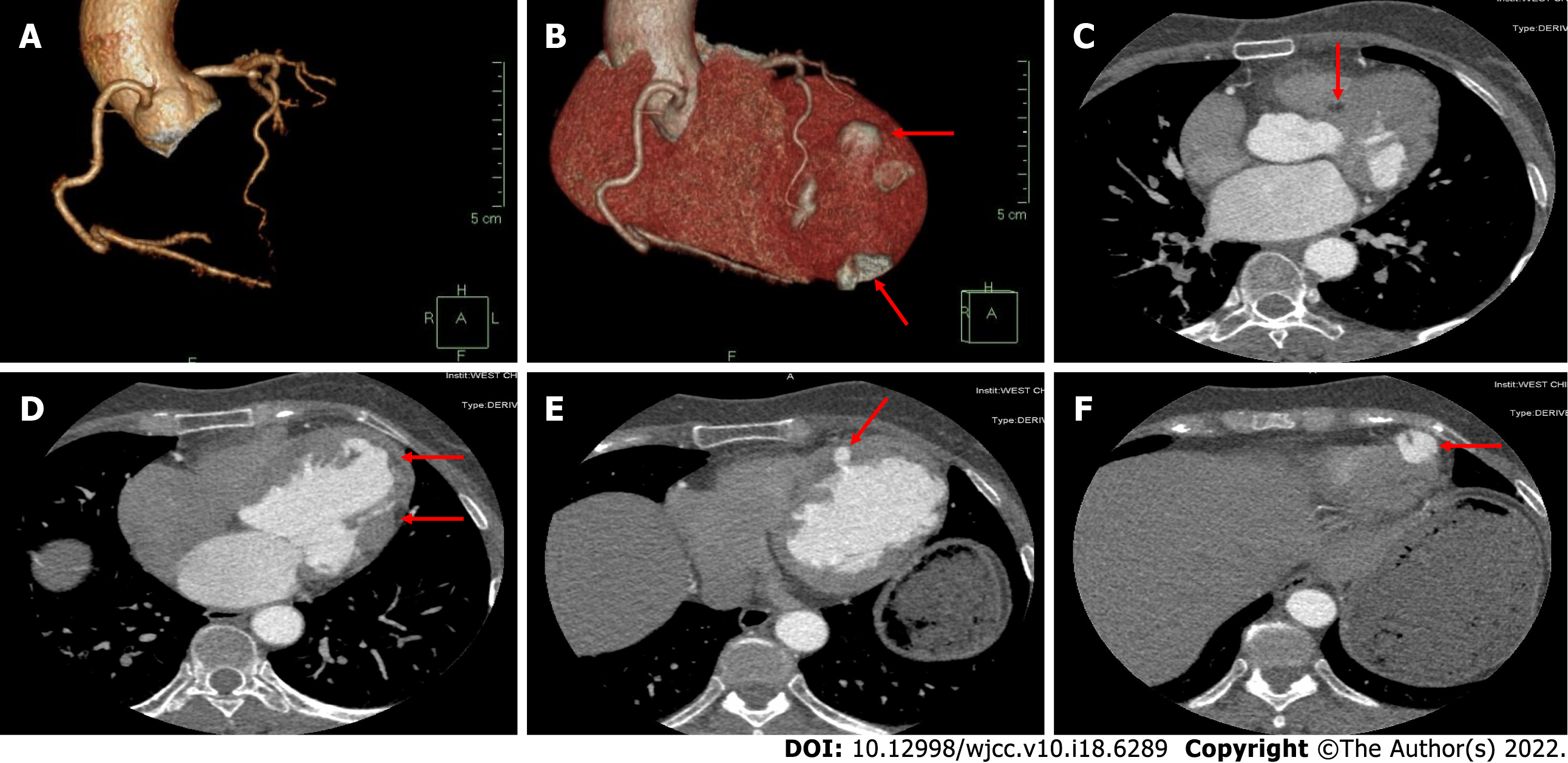
Figure 5 Coronary computerized tomography angiography results of the patient under this study.
A: Three-dimensional reconstruction of coronary arteries and right dominant coronary artery circulation showed no obvious stenosis; B: Three-dimensional reconstruction of the heart showed multiple outpouchings on the left ventricular wall; C: The left ventricular septum myocardium exhibited uneven enhancement. The degree of enhancement is shown by the red arrow and was lower than that of the surrounding tissue, computed tomography value -90 HU; D: Uneven enhancement in the left ventricle free wall myocardium. The degree of enhancement is shown by the red arrow and was lower than that of the surrounding tissue, computed tomography value -114 HU; E: The left ventricular wall exhibited a disordered structure. The uneven thickness of the ventricular wall and local diverticulum is denoted by the red arrow; F: Diverticulum in the apex of the left ventricle is shown by the red arrow.
- Citation: Zhang X, Ye RY, Chen XP. Dilated left ventricle with multiple outpouchings — a severe congenital ventricular diverticulum or left-dominant arrhythmogenic cardiomyopathy: A case report. World J Clin Cases 2022; 10(18): 6289-6297
- URL: https://www.wjgnet.com/2307-8960/full/v10/i18/6289.htm
- DOI: https://dx.doi.org/10.12998/wjcc.v10.i18.6289


