Copyright
©The Author(s) 2022.
World J Clin Cases. Jun 26, 2022; 10(18): 6289-6297
Published online Jun 26, 2022. doi: 10.12998/wjcc.v10.i18.6289
Published online Jun 26, 2022. doi: 10.12998/wjcc.v10.i18.6289
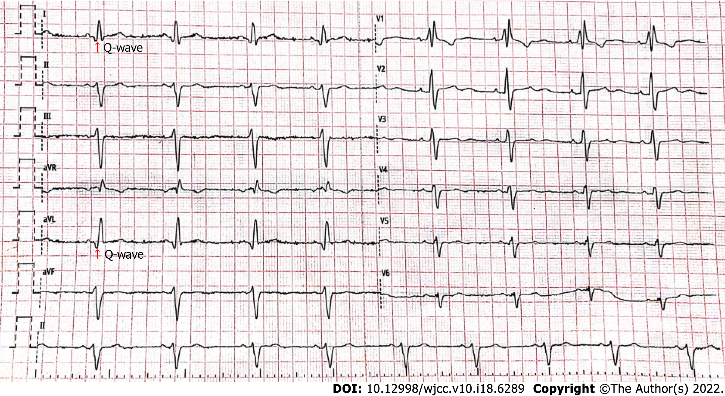
Figure 1 Routine 12-leads electrocardiogram results of the patient under this study.
Electrocardiogram showed left anterior branch block, complete right bundle branch block, high sidewall (I, AVL) abnormal Q wave, and left chest leads low voltage (V4-6) with poor R wave progression.
- Citation: Zhang X, Ye RY, Chen XP. Dilated left ventricle with multiple outpouchings — a severe congenital ventricular diverticulum or left-dominant arrhythmogenic cardiomyopathy: A case report. World J Clin Cases 2022; 10(18): 6289-6297
- URL: https://www.wjgnet.com/2307-8960/full/v10/i18/6289.htm
- DOI: https://dx.doi.org/10.12998/wjcc.v10.i18.6289


