Copyright
©The Author(s) 2022.
World J Clin Cases. Jun 26, 2022; 10(18): 6269-6276
Published online Jun 26, 2022. doi: 10.12998/wjcc.v10.i18.6269
Published online Jun 26, 2022. doi: 10.12998/wjcc.v10.i18.6269
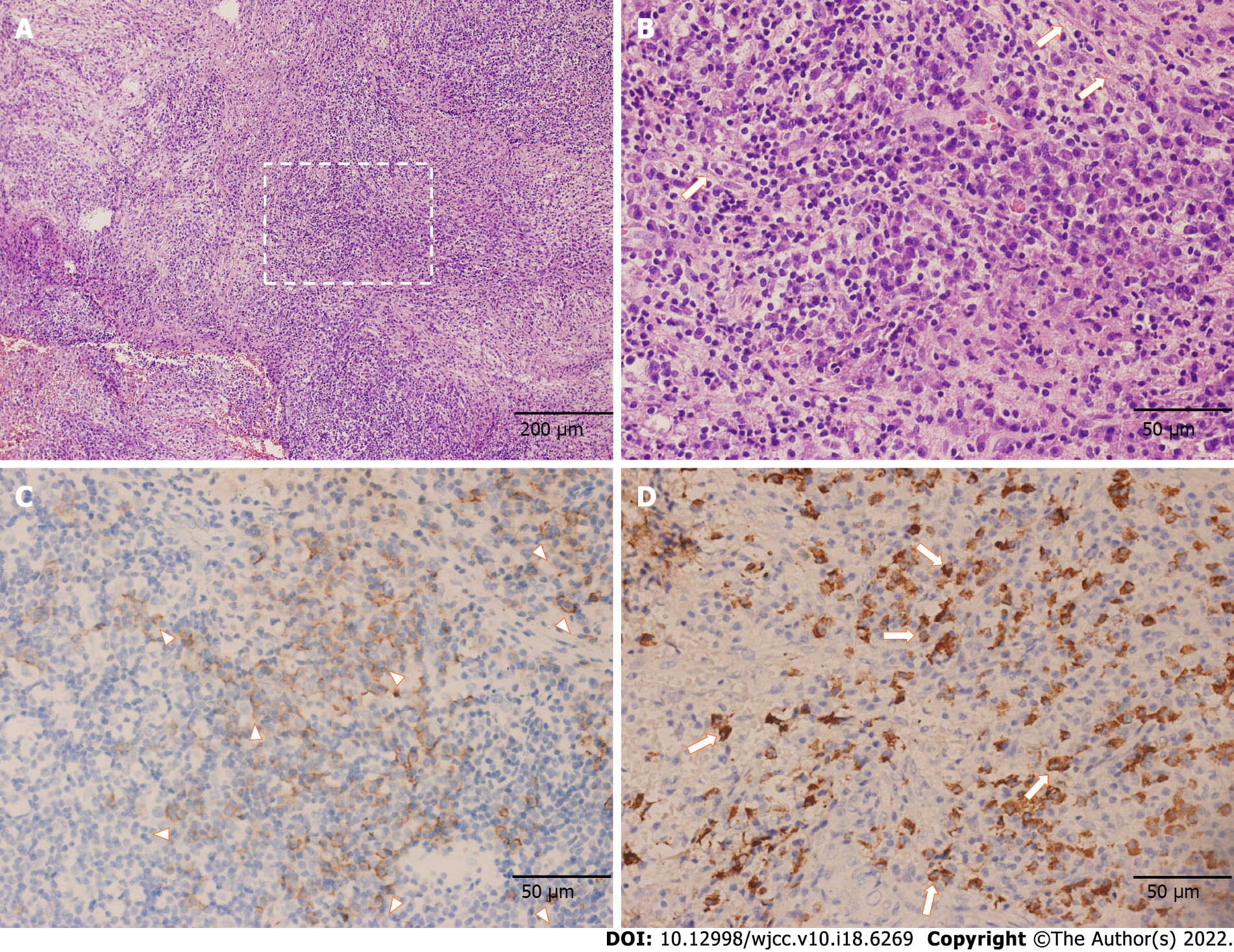
Figure 3 Pathological features of the resection part indicating the diagnosis of immunoglobulin G4 related hypertrophic pachymeningitis.
A and B: Proliferation of fibrous tissue accompanied by numerous lymphocytes and plasma cells as shown by hematoxylin and eosin staining (A: × 100; B: × 400); C: Immunohistochemical staining exhibited an increased number of CD138-positive plasma cells (× 400); D: Large number of immunoglobulin G4-positive plasma cells as shown by immunohistochemical staining [~200 per high power field (× 400)].
- Citation: Yu Y, Lv L, Yin SL, Chen C, Jiang S, Zhou PZ. Clivus-involved immunoglobulin G4 related hypertrophic pachymeningitis mimicking meningioma: A case report. World J Clin Cases 2022; 10(18): 6269-6276
- URL: https://www.wjgnet.com/2307-8960/full/v10/i18/6269.htm
- DOI: https://dx.doi.org/10.12998/wjcc.v10.i18.6269


