Copyright
©The Author(s) 2022.
World J Clin Cases. Jun 26, 2022; 10(18): 6261-6268
Published online Jun 26, 2022. doi: 10.12998/wjcc.v10.i18.6261
Published online Jun 26, 2022. doi: 10.12998/wjcc.v10.i18.6261
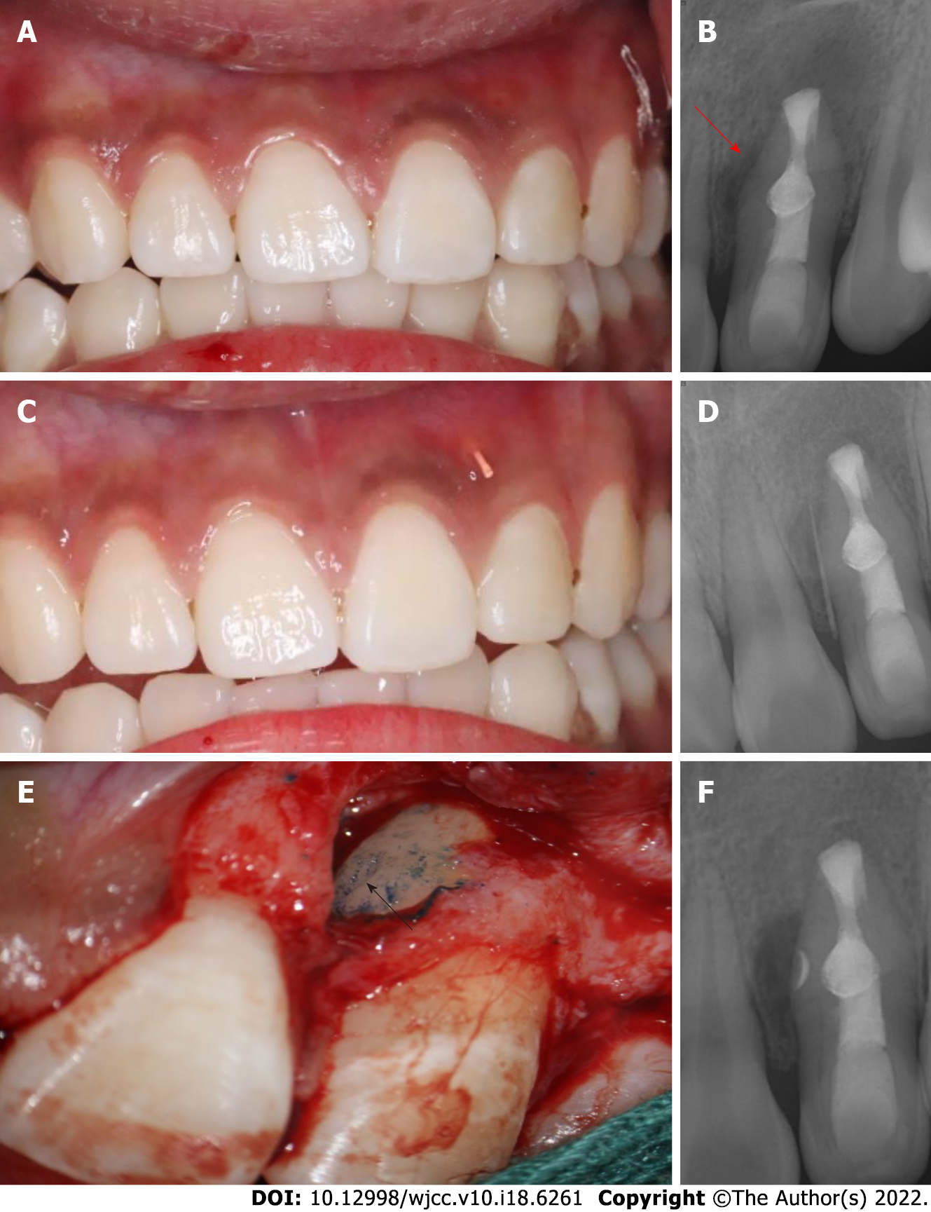
Figure 3 Intraoral examination after 6 mo and surgical treatment.
A: Labial view showing the left lateral incisor with a sinus on the mesiobuccal aspect; B: Radiograph image showing enlargement of a large periapical radiolucency (red arrow) around the mid-root of tooth 22; C: A gutta percha was placed into the sinus of tooth 22; D: Radiograph of tooth 22 with a gutta percha point in the sinus tract; E: Surgical confirmation of the L-shaped pit on the mid-root surface (black arrow); F: Postoperative radiograph.
- Citation: Zhang J, Li N, Li WL, Zheng XY, Li S. Management of type IIIb dens invaginatus using a combination of root canal treatment, intentional replantation, and surgical therapy: A case report. World J Clin Cases 2022; 10(18): 6261-6268
- URL: https://www.wjgnet.com/2307-8960/full/v10/i18/6261.htm
- DOI: https://dx.doi.org/10.12998/wjcc.v10.i18.6261


