Copyright
©The Author(s) 2022.
World J Clin Cases. Jun 26, 2022; 10(18): 6192-6197
Published online Jun 26, 2022. doi: 10.12998/wjcc.v10.i18.6192
Published online Jun 26, 2022. doi: 10.12998/wjcc.v10.i18.6192
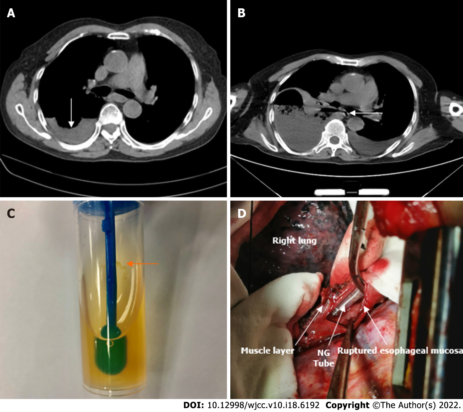
Figure 1 Computed tomography.
A: The chest computed tomography (CT) scan showed a small amount of fluid in the right pleural cavity; B: Chest CT was repeated: right-sided pleural effusion and perforation of the upper left esophagus were observed; C: Grapefruit-like residue drainage liquid was seen; D: The surgery showed the ruptured esophagus with nasogastric tube (NG tube) exposure.
- Citation: Tan N, Luo YH, Li GC, Chen YL, Tan W, Xiang YH, Ge L, Yao D, Zhang MH. Presentation of Boerhaave’s syndrome as an upper-esophageal perforation associated with a right-sided pleural effusion: A case report. World J Clin Cases 2022; 10(18): 6192-6197
- URL: https://www.wjgnet.com/2307-8960/full/v10/i18/6192.htm
- DOI: https://dx.doi.org/10.12998/wjcc.v10.i18.6192


