Copyright
©The Author(s) 2022.
World J Clin Cases. Jun 26, 2022; 10(18): 6175-6183
Published online Jun 26, 2022. doi: 10.12998/wjcc.v10.i18.6175
Published online Jun 26, 2022. doi: 10.12998/wjcc.v10.i18.6175
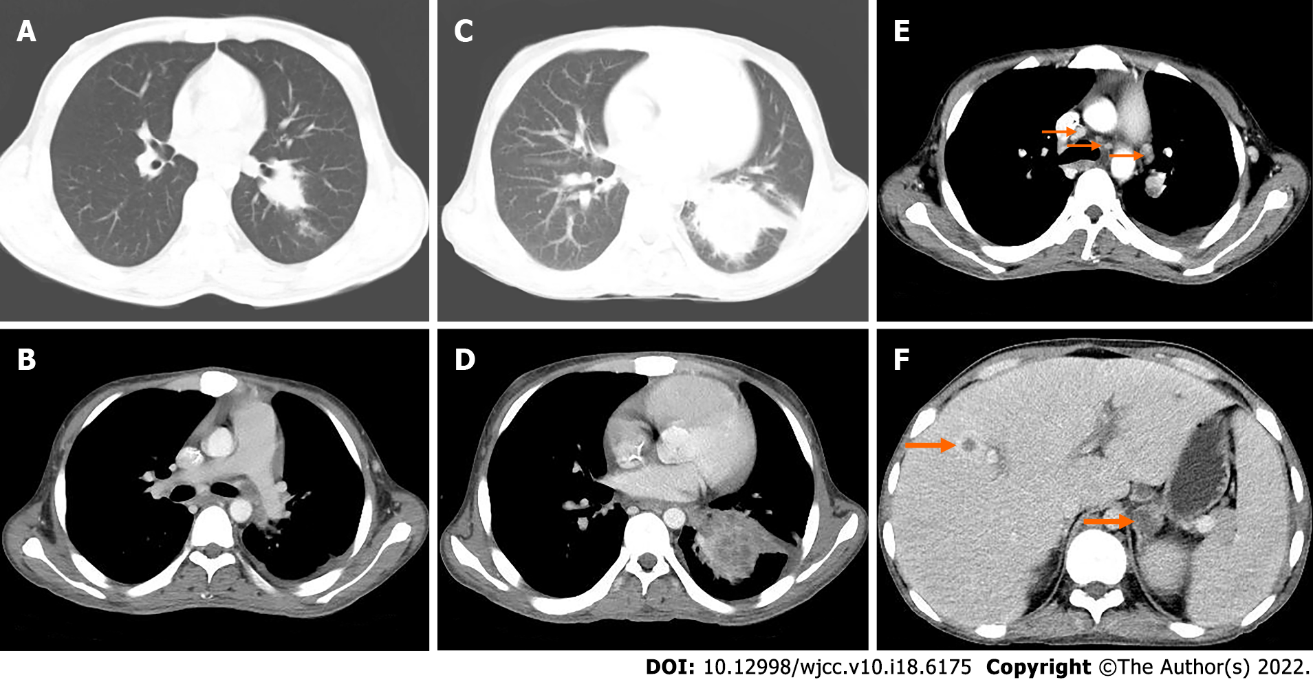
Figure 6 Computed tomography 43 mo (A-B) and 50 mo (C-F) after surgery.
A: The nodule of the lower lobe of the left lung was obviously enlarged, and the boundary was not clear. The size was about 2.5 cm × 3.5 cm; B: The soft tissue nodule of the left pleura showed slight enhancement, with a small amount of fluid density in the left pleural cavity; C: The irregular mass in the lower lobe of the left lung was significantly larger than before, with a size of about 8.2 cm × 5.7 cm; D: After enhancement, the focus showed moderate enhancement; E: Multiple enlarged lymph nodes in mediastinum, left hilum and left axilla; F: Low-density soft tissue mass of the left adrenal gland with unclear boundary and moderate circular enhancement.
- Citation: Huang WP, Li LM, Gao JB. Postoperative multiple metastasis of clear cell sarcoma-like tumor of the gastrointestinal tract in adolescent: A case report. World J Clin Cases 2022; 10(18): 6175-6183
- URL: https://www.wjgnet.com/2307-8960/full/v10/i18/6175.htm
- DOI: https://dx.doi.org/10.12998/wjcc.v10.i18.6175


