Copyright
©The Author(s) 2022.
World J Clin Cases. Jun 26, 2022; 10(18): 6175-6183
Published online Jun 26, 2022. doi: 10.12998/wjcc.v10.i18.6175
Published online Jun 26, 2022. doi: 10.12998/wjcc.v10.i18.6175
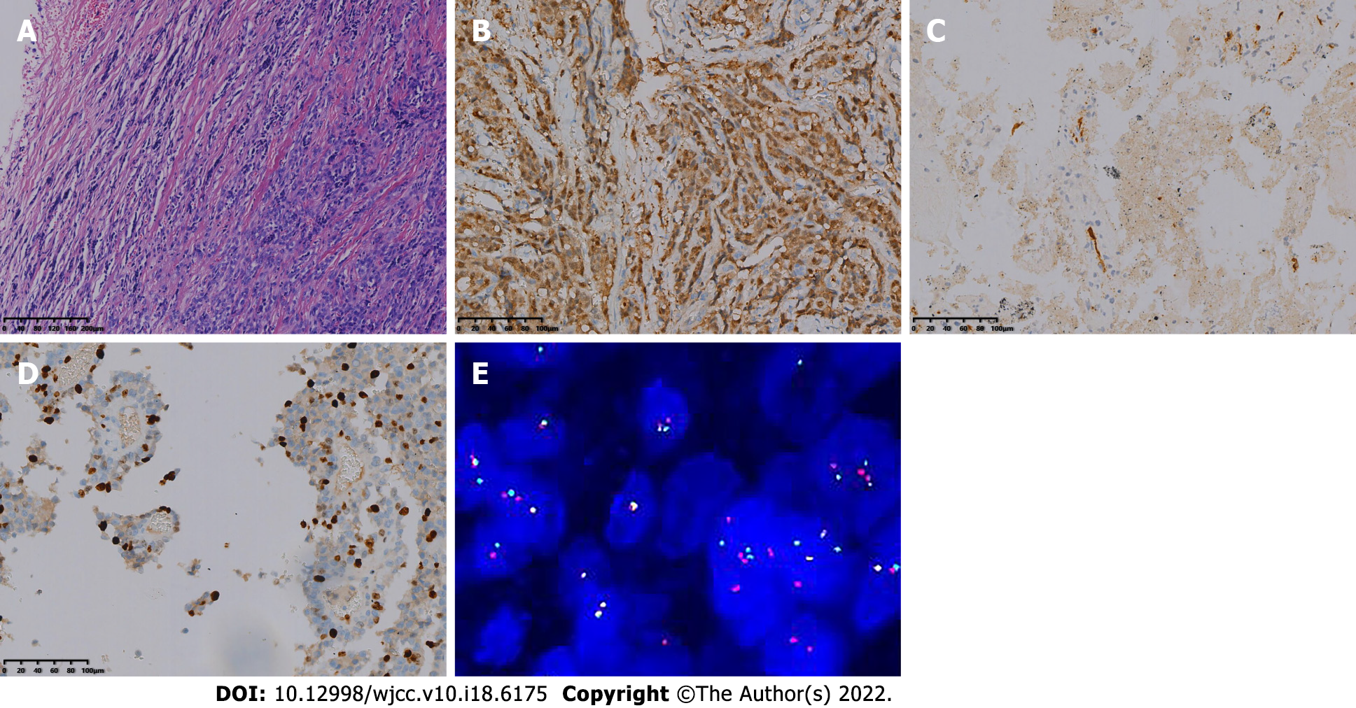
Figure 3 Optical microscopy and molecular pathology fluorescence in situ hybridization.
A: The tumor cells were diffusely arranged, separated by a slender fibrous diaphragm [hematoxylin and eosin (HE) 200×]; B: Immunohistochemical staining revealed S-100 positivity (Envision, 200×); C: Immunohistochemical staining revealed CD34 positivity (Envision, 200×); D: Immunohistochemical staining revealed 30% positivity for Ki-67 (Envision, 200×); E: total of 100 tumor cells were detected and counted, and the number of positive cells was 46%. EWSR1 gene breakage occurred in this case.
- Citation: Huang WP, Li LM, Gao JB. Postoperative multiple metastasis of clear cell sarcoma-like tumor of the gastrointestinal tract in adolescent: A case report. World J Clin Cases 2022; 10(18): 6175-6183
- URL: https://www.wjgnet.com/2307-8960/full/v10/i18/6175.htm
- DOI: https://dx.doi.org/10.12998/wjcc.v10.i18.6175


