Copyright
©The Author(s) 2022.
World J Clin Cases. Jun 26, 2022; 10(18): 6175-6183
Published online Jun 26, 2022. doi: 10.12998/wjcc.v10.i18.6175
Published online Jun 26, 2022. doi: 10.12998/wjcc.v10.i18.6175
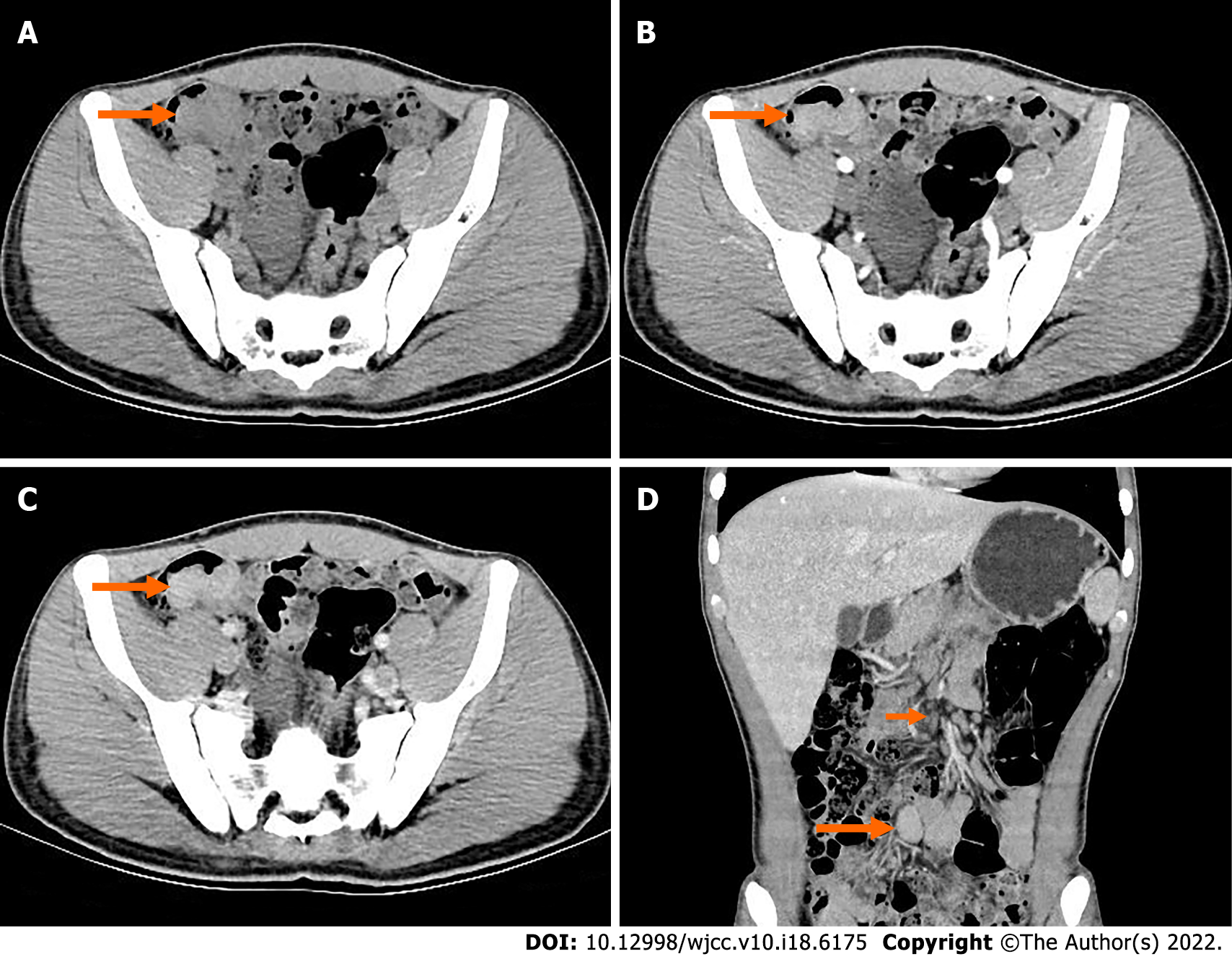
Figure 2 Preoperative computed tomography.
A: Obvious localized thickening of ileal wall in the right lower abdomen; B: The mass showed mild enhancement in the arterial phase; C: The mass showed moderate homogeneous progressive enhancement during the venous phase; D: The venous coronal images showed that the mass of the intestinal wall grew into the intestinal cavity; the intestinal cavity was obviously narrowed; and the lymph nodes at the root of the mesentery were enlarged.
- Citation: Huang WP, Li LM, Gao JB. Postoperative multiple metastasis of clear cell sarcoma-like tumor of the gastrointestinal tract in adolescent: A case report. World J Clin Cases 2022; 10(18): 6175-6183
- URL: https://www.wjgnet.com/2307-8960/full/v10/i18/6175.htm
- DOI: https://dx.doi.org/10.12998/wjcc.v10.i18.6175


