Copyright
©The Author(s) 2022.
World J Clin Cases. Jun 26, 2022; 10(18): 6168-6174
Published online Jun 26, 2022. doi: 10.12998/wjcc.v10.i18.6168
Published online Jun 26, 2022. doi: 10.12998/wjcc.v10.i18.6168
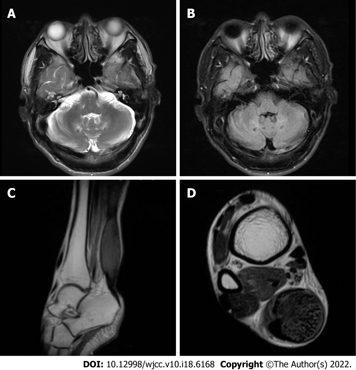
Figure 2 Magnetic resonance imaging.
A, B: Magnetic resonance imaging (MRI) of the brain showed T2-weighted and FLAIR imaging hyperintensity in the bilateral cerebellar dentate nuclei; C, D: MRI of the right ankle showed fusiform swelling and abnormal signals in the achilles tendons.
- Citation: Li ZR, Zhou YL, Jin Q, Xie YY, Meng HM. CYP27A1 mutation in a case of cerebrotendinous xanthomatosis: A case report. World J Clin Cases 2022; 10(18): 6168-6174
- URL: https://www.wjgnet.com/2307-8960/full/v10/i18/6168.htm
- DOI: https://dx.doi.org/10.12998/wjcc.v10.i18.6168


