Copyright
©The Author(s) 2022.
World J Clin Cases. Jun 26, 2022; 10(18): 6050-6059
Published online Jun 26, 2022. doi: 10.12998/wjcc.v10.i18.6050
Published online Jun 26, 2022. doi: 10.12998/wjcc.v10.i18.6050
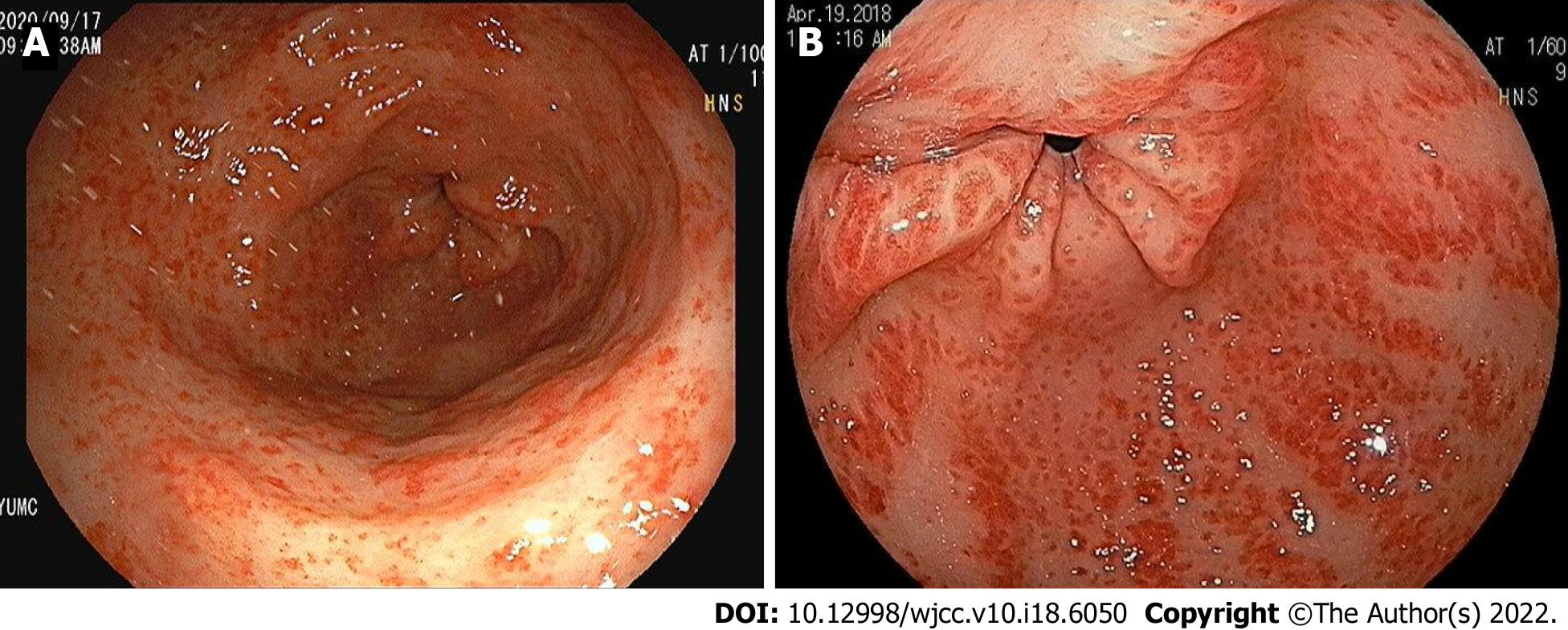
Figure 1 Two main typical endoscopic views of gastric antral vascular ectasia.
A: Punctate (diffuse, honeycomb)-type gastric antral vascular ectasia (GAVE) showing sharply demarcated, punctate red spots diffusely scattered in antrum; B: Striped (linear, watermelon)-type GAVE showing bright red bands radiating longitudinally from pylorus.
- Citation: Kwon HJ, Lee SH, Cho JH. Influences of etiology and endoscopic appearance on the long-term outcomes of gastric antral vascular ectasia. World J Clin Cases 2022; 10(18): 6050-6059
- URL: https://www.wjgnet.com/2307-8960/full/v10/i18/6050.htm
- DOI: https://dx.doi.org/10.12998/wjcc.v10.i18.6050


