Copyright
©The Author(s) 2022.
World J Clin Cases. Jun 26, 2022; 10(18): 6009-6020
Published online Jun 26, 2022. doi: 10.12998/wjcc.v10.i18.6009
Published online Jun 26, 2022. doi: 10.12998/wjcc.v10.i18.6009
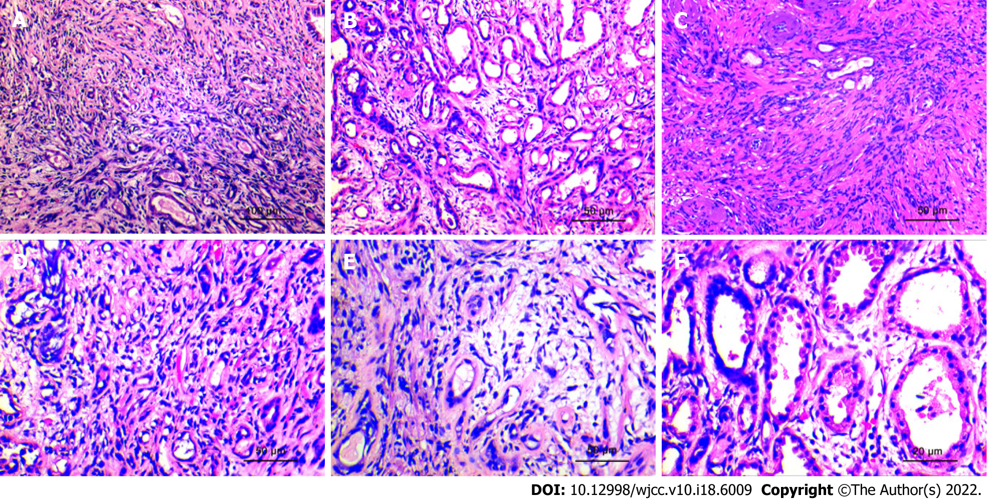
Figure 3 Histopathological morphological characteristics.
A: The lesions are distributed uniformly, have a nodular shape, without obvious capsules, and the simulated prostate adenocarcinoma presents an "invasive growth" pattern. (HE × 100; scale bar, 100 μm); B: The hyperplastic stroma squeezes the glands to form glandular tubes of varying sizes. Eosinophilic shoed spike-like protrusions can be seen in part of the lumen (HE × 200; scale bar, 50 μm); C: The glands of sclerosing adenopathy are squeezed into a cord-like, thread-like, or even single-cell-like arrangement, and the cells have a shuttle-like shape (HE × 200; scale bar, 50 μm); D: When the glandular transition is squeezed, it can form a vacuole-like structure, and the increase in the number of vacuole-like cells leads to a signet ring-like morphology (HE × 200; scale bar, 50 μm); E: The interstitium of the disease was composed of mildly morphological spindle cells, often with collagenous or mucinous changes (HE × 200; scale bar, 50 μm); F: At high power, the glandular epithelium and the compressed myoepithelial components can be seen, the cell cytoplasm is eosinophilic, and the nucleus is located at the base (HE × 400; scale bar, 20 μm).
- Citation: Feng RL, Tao YP, Tan ZY, Fu S, Wang HF. Prostate sclerosing adenopathy: A clinicopathological and immunohistochemical study of twelve patients. World J Clin Cases 2022; 10(18): 6009-6020
- URL: https://www.wjgnet.com/2307-8960/full/v10/i18/6009.htm
- DOI: https://dx.doi.org/10.12998/wjcc.v10.i18.6009


