Copyright
©The Author(s) 2022.
World J Clin Cases. Jun 26, 2022; 10(18): 6009-6020
Published online Jun 26, 2022. doi: 10.12998/wjcc.v10.i18.6009
Published online Jun 26, 2022. doi: 10.12998/wjcc.v10.i18.6009
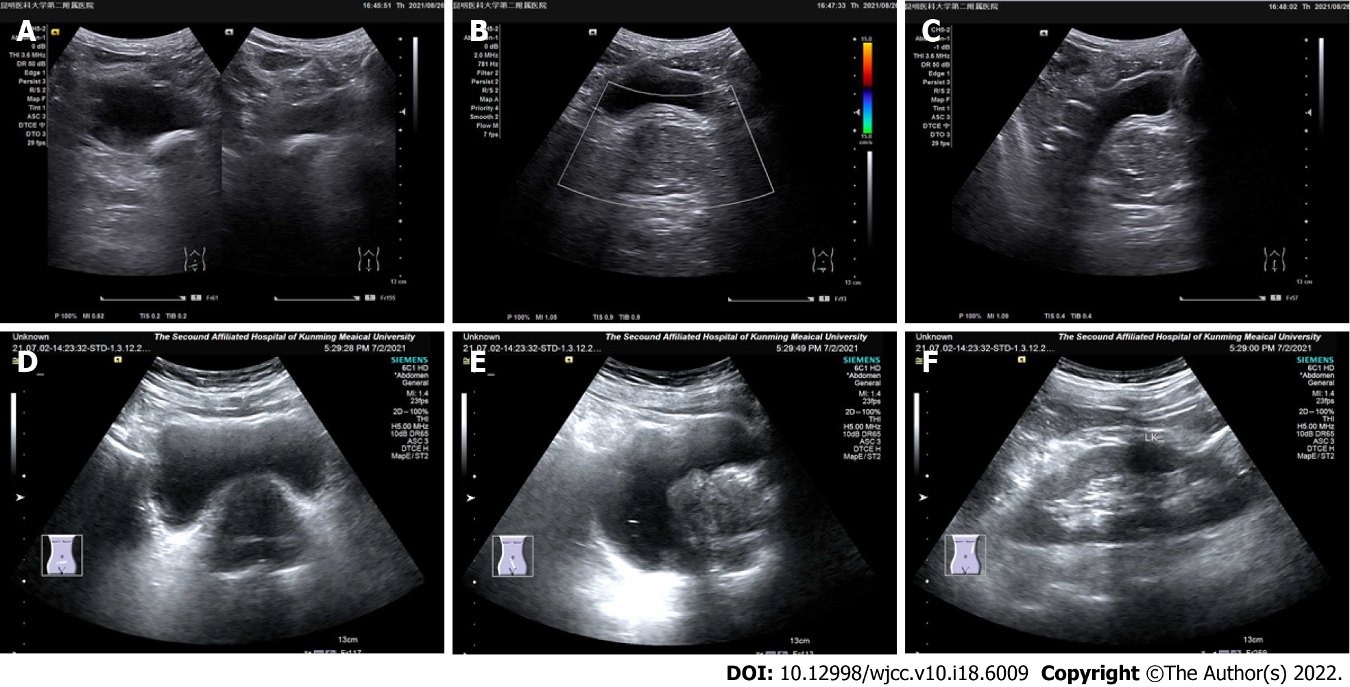
Figure 2 Ultrasound image of sclerosing adenopathy of the prostate.
A-C: The prostate shape was full, showing regular margins, a normal ratio of internal to external glands, an uneven echo, and a sonographic image of benign prostatic hyperplasia; D-F: The prostate gland was enlarged, its shape was plump, the internal gland was enlarged, the external gland was compressed and thinned, the parenchymal echo was not uniform, and the parenchyma was probed with multiple hyperechoic spots.
- Citation: Feng RL, Tao YP, Tan ZY, Fu S, Wang HF. Prostate sclerosing adenopathy: A clinicopathological and immunohistochemical study of twelve patients. World J Clin Cases 2022; 10(18): 6009-6020
- URL: https://www.wjgnet.com/2307-8960/full/v10/i18/6009.htm
- DOI: https://dx.doi.org/10.12998/wjcc.v10.i18.6009


